File:Jenkins-table03.jpg
From Embryology
Size of this preview: 567 × 599 pixels. Other resolution: 992 × 1,048 pixels.
Original file (992 × 1,048 pixels, file size: 293 KB, MIME type: image/jpeg)
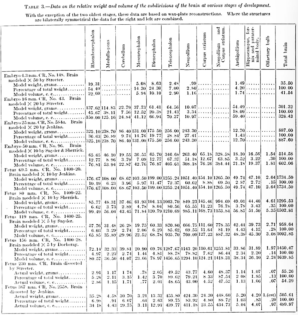
Table 3. Data on the relative weight and volume of the subdivisions of the brain at various stages of development
With the exception of the two oldest stages, these data are based on wax-plate reconstructions. Where the structures are bilaterally symmetrical the data for the right and left are combined.
File history
Click on a date/time to view the file as it appeared at that time.
| Date/Time | Thumbnail | Dimensions | User | Comment | |
|---|---|---|---|---|---|
| current | 18:02, 15 February 2011 | | 992 × 1,048 (293 KB) | S8600021 (talk | contribs) | ==Table 3. Data on the relative weight and volume of the subdivisions of the brain at various stages of development== With the exception of the two oldest stages, these data are based on wax-plate reconstructions. Where the structures are bilaterally sym |
You cannot overwrite this file.
File usage
The following page uses this file: