File:Infant ovary.jpg: Difference between revisions
From Embryology
| (7 intermediate revisions by 2 users not shown) | |||
| Line 1: | Line 1: | ||
==Infant Ovary (human)== | ==Infant Ovary (human)== | ||
In this infant ovary there are a large number of primordial follicles ( | In this infant ovary there are a large number of primordial follicles containing immature oocytes ({{primordial germ cell}}) forming a thick cortical region and no later stages follicle development. Compare this structure with the other images of an ovary after puberty with reproductive activity. | ||
* inset - region of whole ovary shown in this image | * inset - region of whole ovary shown in this image | ||
* right - ovary surface (tunica | * right - ovary surface ('''{{tunica albuginea}}''') | ||
* centre - ovary cortex | * centre - ovary '''cortex''' | ||
* left - ovary medulla | * left - ovary '''medulla''' | ||
* '''{{primordial follicle}}''' - primary oocyte is surrounded by a single layer of follicular cells. | |||
* '''oocytes''' - enter diplotene stage of {{meiosis}} prophase. | |||
** remain arrested in this early stage of the '''first meiotic division''' as a primary oocyte (primordial follicle) | |||
[[Category:Ovary]] [[Category:Histology]] [[Category:Genital]] [[Category:Oocyte]] | |||
:'''Links:''' [[Ovary Development]] | [[Oocyte Development]] | {{Meiosis}} | |||
{{footer}} | |||
[[Category:Ovary]] [[Category:Female]] [[Category:Histology]] [[Category:Genital]] [[Category:Oocyte]] | |||
Latest revision as of 08:44, 28 April 2020
Infant Ovary (human)
In this infant ovary there are a large number of primordial follicles containing immature oocytes (primordial germ cell) forming a thick cortical region and no later stages follicle development. Compare this structure with the other images of an ovary after puberty with reproductive activity.
- inset - region of whole ovary shown in this image
- right - ovary surface (tunica albuginea)
- centre - ovary cortex
- left - ovary medulla
- primordial follicle - primary oocyte is surrounded by a single layer of follicular cells.
- oocytes - enter diplotene stage of meiosis prophase.
- remain arrested in this early stage of the first meiotic division as a primary oocyte (primordial follicle)
- Links: Ovary Development | Oocyte Development | meiosis
Cite this page: Hill, M.A. (2024, May 5) Embryology Infant ovary.jpg. Retrieved from https://embryology.med.unsw.edu.au/embryology/index.php/File:Infant_ovary.jpg
- © Dr Mark Hill 2024, UNSW Embryology ISBN: 978 0 7334 2609 4 - UNSW CRICOS Provider Code No. 00098G
File history
Click on a date/time to view the file as it appeared at that time.
| Date/Time | Thumbnail | Dimensions | User | Comment | |
|---|---|---|---|---|---|
| current | 23:12, 21 September 2009 | 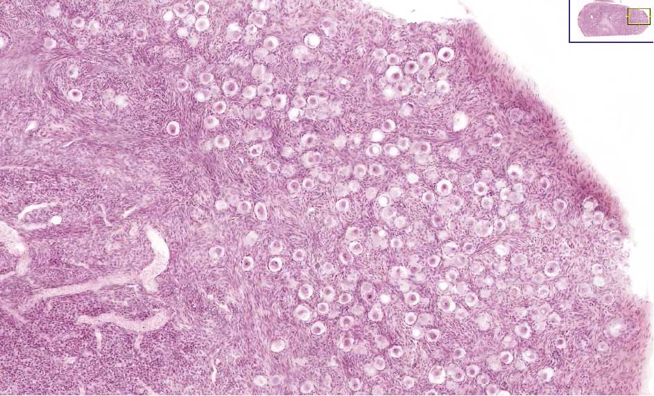 | 943 × 571 (108 KB) | S8600021 (talk | contribs) | Infant Ovary, note the large number of primordial follicles around the cortex. |
You cannot overwrite this file.
File usage
The following 24 pages use this file:
- 2009 Lecture 16
- 2010 BGD Practical 3 - Oogenesis and Ovulation
- 2010 Lecture 16
- 2011 Lab 1 - Oogenesis
- 2011 Lab 8 - Postnatal
- 2011 Lecture 16
- 2014 Group Project 4
- ANAT2341 Lab 1 - Oogenesis
- ANAT2341 Lab 8 - Postnatal
- BGDA Lecture - Development of the Embryo/Fetus 1
- BGDA Practical 3 - Oogenesis and Ovulation
- BGDB Sexual Differentiation - Postnatal
- BGD Lecture - Endocrine Development
- BGD Lecture - Sexual Differentiation
- Endocrine - Gonad Development
- In Vitro Oogenesis
- Lecture - Endocrine Development
- Lecture - Fertilization
- Lecture - Genital Development
- Oocyte Development
- Ovary Development
- REI - Reproductive Medicine Seminar 2018
- Royal Hospital for Women - Reproductive Medicine Seminar 2018
- Talk:2014 Group Project 4