File:Hydatidiform mole 02.jpg: Difference between revisions
mNo edit summary |
m (→Reference) |
||
| Line 12: | Line 12: | ||
===Reference=== | ===Reference=== | ||
<pubmed>19774194</pubmed> | <pubmed>19774194</pubmed> | ||
| Line 24: | Line 19: | ||
This is an open-access article distributed under the terms of the Creative Commons Attribution License, which permits unrestricted use, distribution, and reproduction in any medium, provided the original work is properly cited. | This is an open-access article distributed under the terms of the Creative Commons Attribution License, which permits unrestricted use, distribution, and reproduction in any medium, provided the original work is properly cited. | ||
Indian J Radiol Imaging. 2008 November; 18(4): 326–344. | |||
doi: 10.4103/0971-3026.43848. | |||
Original file name: IJRI-18-326-g013.jpg | |||
[[Category:Placenta]] [[Category:Ultrasound]] [[Category:Human]] [[Category:Abnormal Development]] [[Category:Uterus]] | [[Category:Placenta]] [[Category:Ultrasound]] [[Category:Human]] [[Category:Abnormal Development]] [[Category:Uterus]] | ||
Revision as of 22:51, 10 June 2013
Hydatidiform mole
In this patient with a 6-weeks' gestation presenting with vaginal bleeding, transvaginal USG
(A) shows a gestational sac (white arrow) on the first examination. Follow-up examination after 2 days
(B) shows focal cystic changes (arrow) with loss of normal definition of the gestational sac, suggesting the possibility of molar changes. Investigations confirmed triploidy
- Links: Hydatidiform Mole
Reference
<pubmed>19774194</pubmed>
Copyright
© Indian Journal of Radiology and Imaging
This is an open-access article distributed under the terms of the Creative Commons Attribution License, which permits unrestricted use, distribution, and reproduction in any medium, provided the original work is properly cited.
Indian J Radiol Imaging. 2008 November; 18(4): 326–344. doi: 10.4103/0971-3026.43848.
Original file name: IJRI-18-326-g013.jpg
File history
Click on a date/time to view the file as it appeared at that time.
| Date/Time | Thumbnail | Dimensions | User | Comment | |
|---|---|---|---|---|---|
| current | 02:34, 27 May 2010 | 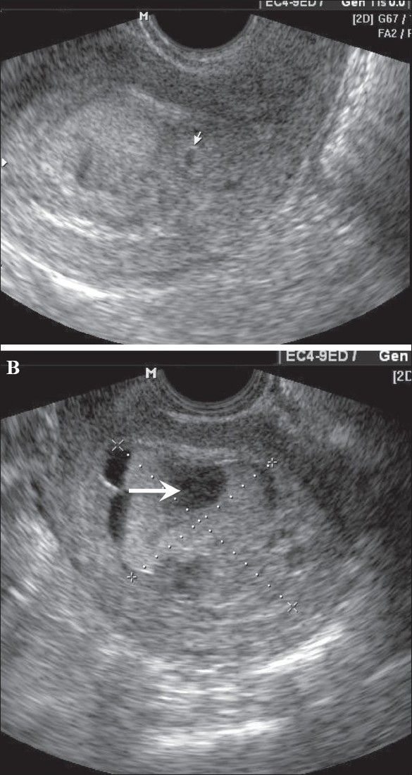 | 585 × 1,100 (237 KB) | S8600021 (talk | contribs) | ==Hydatidiform mole== In this patient with a 6-weeks' gestation presenting with vaginal bleeding, transvaginal USG (A) shows a gestational sac (white arrow) on the first examination. Follow-up examination after 2 days (B) shows focal cystic changes ( |
You cannot overwrite this file.
File usage
The following page uses this file: