File:Huntingtin structure.jpg
Huntingtin_structure.jpg (595 × 266 pixels, file size: 39 KB, MIME type: image/jpeg)
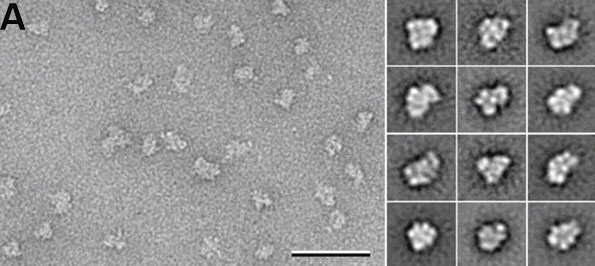
Huntingtin conformational flexibility and domain organization.
(A) Representative area of an electron micrograph of negatively stained FLAG-Q23 huntingtin (scale bar 50 nm) (left) and representative class averages (side length of panels is 28.8 nm) (right) showing the structural variability of huntingtin.
1.
Huntingtin facilitates polycomb repressive complex 2. Seong IS, Woda JM, Song JJ, Lloret A, Abeyrathne PD, Woo CJ, Gregory G, Lee JM, Wheeler VC, Walz T, Kingston RE, Gusella JF, Conlon RA, Macdonald ME. Hum Mol Genet. 2010 Feb 15;19(4):573-83. Epub 2009 Nov 23. PMID: 19933700 | Hum Mol Genet.
Extract from Figure 1. http://hmg.oxfordjournals.org/content/vol19/issue4/images/large/ddp52401.jpeg
© The Author 2009. Published by Oxford University Press
This is an Open Access article distributed under the terms of the Creative Commons Attribution Non-Commercial License (http://creativecommons.org/licenses/by-nc/2.5), which permits unrestricted non-commercial use, distribution, and reproduction in any medium, provided the original work is properly cited.
File history
Click on a date/time to view the file as it appeared at that time.
| Date/Time | Thumbnail | Dimensions | User | Comment | |
|---|---|---|---|---|---|
| current | 12:55, 22 April 2010 |  | 595 × 266 (39 KB) | S8600021 (talk | contribs) | Huntingtin conformational flexibility and domain organization. (A) Representative area of an electron micrograph of negatively stained FLAG-Q23 huntingtin (scale bar 50 nm) (left) and representative class averages (side length of panels is 28.8 nm) (rig |
You cannot overwrite this file.
File usage
There are no pages that use this file.
