File:Human week 10 fetus 11.jpg

Original file (1,200 × 900 pixels, file size: 304 KB, MIME type: image/jpeg)
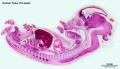
Human Female Fetus - Sacrum (10 week)
Large image version of plane D, close to midline (Stain - Haematoxylin Eosin). 0.3 mm scale bar
Sacral vertebra (S1–S5) and coccyx (last 4 vertebra) both shown unfused and as cartilage, formed from sclerotome of somites 31 to 44. The cartilage will later ossify in the fetal period. The vertebra will later fuse postnatally from adolescence to the adult (18 - 30 years).
Note the remnant of the notochord present in the vertebral body.
Spinal cord (medulla spinalis), conus medullaris, dura mater, film terminale. Spinal cord has developed from the neural tube.
- conus medullaris - (Latin, "medullary cone") is the tapered lower end of the spinal cord.
Rectum lies anterior to the vertebra and has developed from the hindgut. Note the differentiated epithelial wall of the rectum.
- Human Female Fetus (week 10)
Sagittal Section (plane D)

Pituitary and Lamina Terminalis

Olfactory Nerve

Atlas and Axis

Sacrum

Oral Cavity

Epiglottis

Heart

Spleen

Midgut Herniation

Midgut Herniation (label)

Pelvic Region

Pelvic Region (label)


Related Images
Fetus (week 10) Planes A (most lateral), B (lateral), C (medial) and D (midline) from lateral towards the midline.
- Human Fetus - most lateral | lateral | medial | midline
- Head - most lateral | lateral | medial | midline
- Cerebellum - most lateral | lateral | medial | midline
- Urogenital Unlabelled - most lateral | lateral | medial | midline
- Urogenital Labelled - most lateral | lateral | medial | midline
- Large Images - midline
- Image Source: UNSW Embryology, no reproduction without permission.
Cite this page: Hill, M.A. (2024, April 27) Embryology Human week 10 fetus 11.jpg. Retrieved from https://embryology.med.unsw.edu.au/embryology/index.php/File:Human_week_10_fetus_11.jpg
- © Dr Mark Hill 2024, UNSW Embryology ISBN: 978 0 7334 2609 4 - UNSW CRICOS Provider Code No. 00098G
File history
Click on a date/time to view the file as it appeared at that time.
| Date/Time | Thumbnail | Dimensions | User | Comment | |
|---|---|---|---|---|---|
| current | 23:14, 17 June 2012 | | 1,200 × 900 (304 KB) | Z8600021 (talk | contribs) |
You cannot overwrite this file.
File usage
The following 18 pages use this file:
- BGDA Practical 12 - Embryo to Fetus
- Fetal Development - 10 Weeks
- Foundations Practical - Week 9 to 36
- Neural - Spinal Cord Development
- File:Human week 10 fetus 01.jpg
- File:Human week 10 fetus 03.jpg
- File:Human week 10 fetus 04.jpg
- File:Human week 10 fetus 05.jpg
- File:Human week 10 fetus 06.jpg
- File:Human week 10 fetus 07.jpg
- File:Human week 10 fetus 08.jpg
- File:Human week 10 fetus 09.jpg
- File:Human week 10 fetus 10.jpg
- File:Human week 10 fetus 11.jpg
- File:Human week 10 fetus 12.jpg
- File:Human week 10 fetus 23.jpg
- File:Human week 10 fetus 26.jpg
- Template:Human Female Fetus Week 10 gallery