File:Human placenta SERPINE2 expression 01.jpg

Original file (800 × 601 pixels, file size: 134 KB, MIME type: image/jpeg)
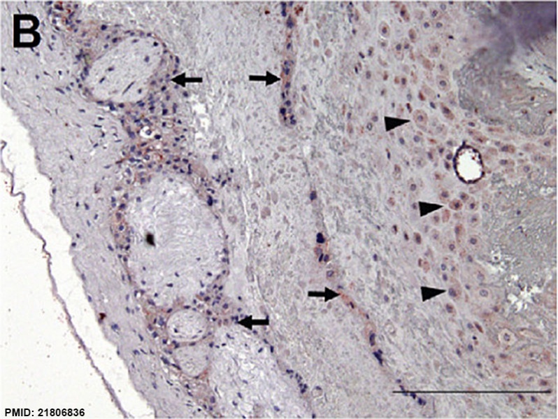
Human placenta SERPINE2 expression
(B) Moderate staining of extravillous trophoblasts (arrow) and decidual cells (arrowhead) in the chorionic plate, and very low staining in the chorionic mesoderm and fibrinoid deposits.
Scale bars represent 200 μm
(A) Expression of SERPINE2 mRNA in human placentas in different trimesters. Levels of SERPINE2 mRNA were determined by real-time quantitative PCR in 19 placental tissues collected in three gestational trimesters (T1, T2, and T3; n = 5, 4, and 10, respectively). For each sample, the SERPINE2 expression level was normalized to the expression level of the RPLPO gene in the same sample. Data are presented as the mean ± SEM. * p < 0.05. Immunohistochemical staining showed the distribution of the SERPINE2 protein in the human placenta at gestational week 15. (B) Moderate staining of extravillous trophoblasts (arrow) and decidual cells (arrowhead) in the chorionic plate, and very low staining in the chorionic mesoderm and fibrinoid deposits. (C) Positive immunostaining was extensively detected in decidual cells (dc), cytotrophoblasts, extravillous trophoblasts at the junction zone of the cell column (cc) and anchoring villi (av), and the endothelia of the spiral artery (sa); and weak staining was found in fibrinoids (f) and the villous mesenchyme. (D) The dashed-lined region in 1C was magnified to show intense staining in syncytiotrophoblasts and cytotrophoblasts in floating villi. The invaded extravillous trophoblasts (arrow) and decidual cells (arrowhead) at the basal plate were strongly stained. (E) Positive staining of cytokeratin (CK)-7 was confirmed in syncytiotrophoblasts and cytotrophoblasts in floating villi, and invading trophoblasts (arrow). (F) Upper panels, dashed-lined rectangle regions in D and E were magnified to show strong staining of SERPINE2 (left) and CK-7 (right) in syncytiotrophoblasts (st) and cytotrophoblasts (ct) in floating villi. Lower panels, most of the endothelia of spiral arteries were positively stained with anti-SERPINE2 (left), and anti-CK-7 (right) antibodies. (G) Negative staining of control antiserum. Scale bars represent 200 μm (B, C), and 50 μm (D, E, G).
Reference
Chern et al. Reproductive Biology and Endocrinology 2011 9:106 doi:10.1186/1477-7827-9-106
1477-7827-9-106-1-l.jpg
File history
Click on a date/time to view the file as it appeared at that time.
| Date/Time | Thumbnail | Dimensions | User | Comment | |
|---|---|---|---|---|---|
| current | 17:26, 9 June 2014 |  | 800 × 601 (134 KB) | Z8600021 (talk | contribs) | ==Human placenta SERPINE2 expression== (B) Moderate staining of extravillous trophoblasts (arrow) and decidual cells (arrowhead) in the chorionic plate, and very low staining in the chorionic mesoderm and fibrinoid deposits. Scale bars represent 200... |
You cannot overwrite this file.
File usage
The following 2 pages use this file: