File:Human ovulation 01.jpg: Difference between revisions
No edit summary |
|||
| Line 1: | Line 1: | ||
==Human Ovulation== | ==Human Ovulation== | ||
Laparoscopic observation of spontaneous human ovulation. A remarkably prominent vascular pattern was observed on the mature follicle (F, white arrows). A small follicular area called the stigma (S) was seen protruding like a reddish bleb from the follicular surface, with viscous yellow fluid (black arrows) evaginating outward into the peritoneal cavity. The viscous fluid probably carried with it the cumulus–oocyte complex, surrounded by several thousand small granulosa cells known as corona radiata. | Laparoscopic observation of spontaneous human ovulation. A remarkably prominent vascular pattern was observed on the mature follicle (F, white arrows). | ||
A small follicular area called the stigma (S) was seen protruding like a reddish bleb from the follicular surface, with viscous yellow fluid (black arrows) evaginating outward into the peritoneal cavity. | |||
The viscous fluid probably carried with it the cumulus–oocyte complex, surrounded by several thousand small granulosa cells known as corona radiata. | |||
'''Legend''' | '''Legend''' | ||
Revision as of 14:46, 9 December 2012
Human Ovulation
Laparoscopic observation of spontaneous human ovulation. A remarkably prominent vascular pattern was observed on the mature follicle (F, white arrows).
A small follicular area called the stigma (S) was seen protruding like a reddish bleb from the follicular surface, with viscous yellow fluid (black arrows) evaginating outward into the peritoneal cavity.
The viscous fluid probably carried with it the cumulus–oocyte complex, surrounded by several thousand small granulosa cells known as corona radiata.
Legend
- F - mature follicle, white arrows
- S - stigma
- black arrows - ovulating follicular fluid containing cumulus–oocyte surrounded by several thousand small granulosa cells
- Links: Full figure | Image 1 of 4 | Image 2 of 4 | Image 3 of 4 | Image 4 of 4 | Non-ovulating Ovary Image | Ovary Development | Uterus Development | Menstrual Cycle
Original file name: Figure 1
Reference
<pubmed>18440526</pubmed>
Special thanks to Prof. Jacques Donnez.
Fertility and Sterility http://www.fertstert.org/
Science Direct http://www.sciencedirect.com/science/journal/00150282
- “Reprinted from Fertility and Sterility, Sep;90(3), Lousse JC, Donnez J., Laparoscopic observation of spontaneous human ovulation., 833-4., Copyright (2008), with permission from Elsevier.”
License number 2562880840005
File history
Click on a date/time to view the file as it appeared at that time.
| Date/Time | Thumbnail | Dimensions | User | Comment | |
|---|---|---|---|---|---|
| current | 16:40, 6 December 2010 | 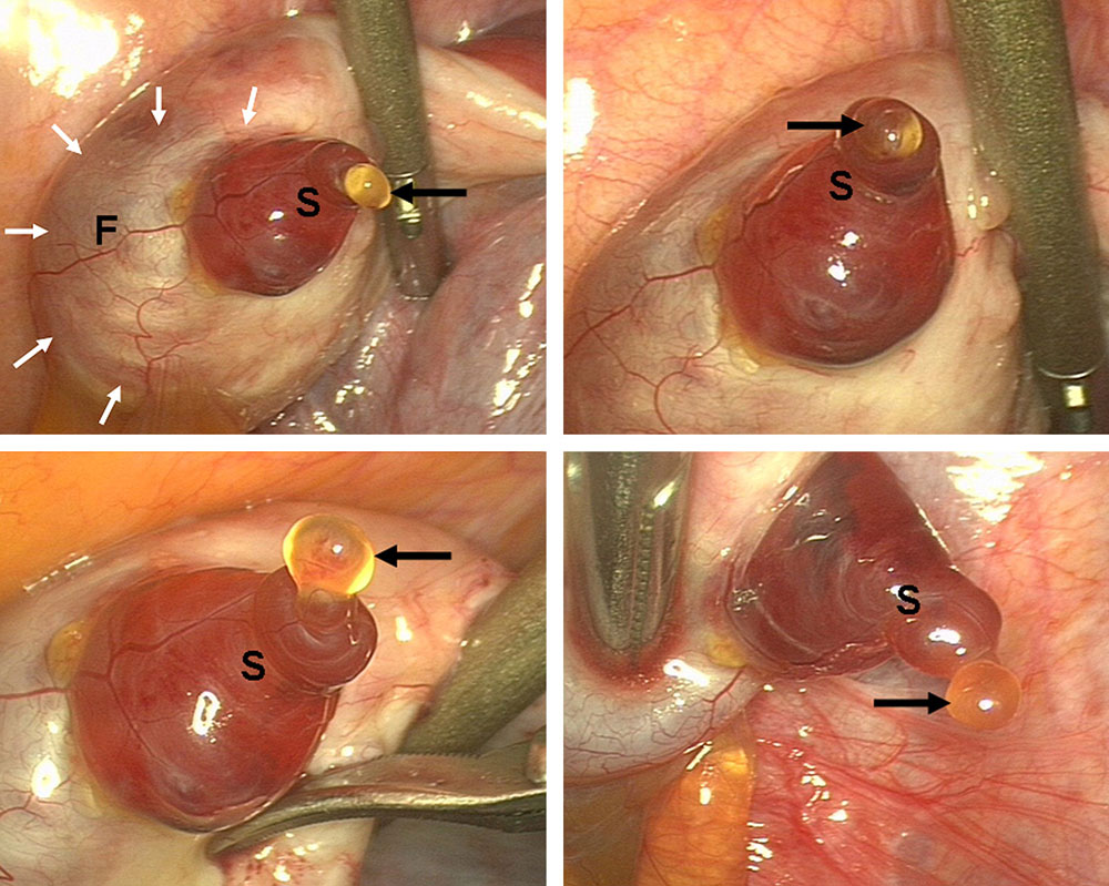 | 1,000 × 799 (175 KB) | S8600021 (talk | contribs) | ==Human Ovulation== Laparoscopic observation of spontaneous human ovulation. A remarkably prominent vascular pattern was observed on the mature follicle (F, white arrows). A small follicular area called the stigma (S) was seen protruding like a reddish b |
You cannot overwrite this file.