File:Human ovary postnatal growth.jpg
From Embryology
No higher resolution available.
Human_ovary_postnatal_growth.jpg (800 × 467 pixels, file size: 40 KB, MIME type: image/jpeg)
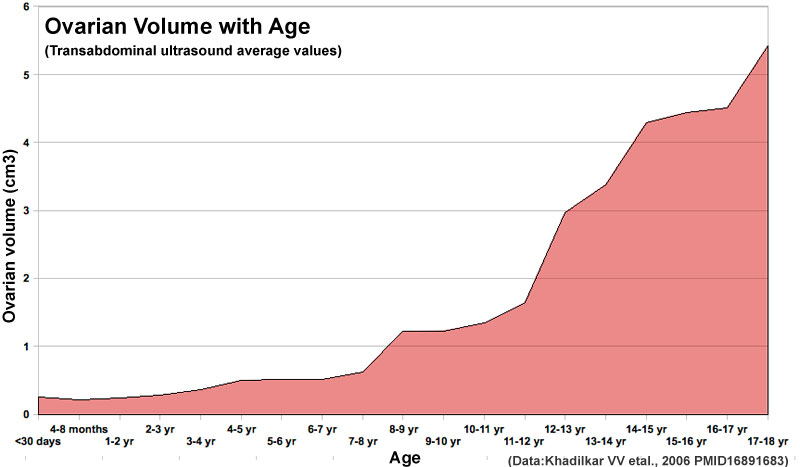
Human Ovary Postnatal Growth
Graph was constructed from Indian population data shown in "TABLE I – Ovarian Volume, Uterine Length and FCR According to Chronological Age (n = 214)"
- "The aim of our study was to determine the pattern of female reproductive organ growth in Indian girls from birth to 18 years of age and to correlate the uterine length, mean ovarian volume (MOV) and Fundo Cervical Ratio (FCR) with chronological age, bone age and pubertal breast staging. A cross sectional study was performed on 218 girls from birth to 18 years of age. Height, weight, stage of puberty, X-ray for bone age and transabdominal ultrasounds were performed on all girls. Higher chronological age, bone age and increase in breast stage significantly predicted higher MOV (P < 0.001) and higher uterine length (P < 0.001). The MOV, uterine length and FCR are positively correlated with chronological age, bone age, height, weight and breast staging. Data from present study may be useful in screening cases of precocious puberty and other disorders that may need further evaluation."
See also:
- Brazilian data - PMID12034633 AJR Am J Roentgenol.
- Danish data - PMID8521066
Reference
<pubmed>16891683</pubmed>
File history
Click on a date/time to view the file as it appeared at that time.
| Date/Time | Thumbnail | Dimensions | User | Comment | |
|---|---|---|---|---|---|
| current | 16:41, 4 January 2011 | | 800 × 467 (40 KB) | S8600021 (talk | contribs) | ==Human Ovary Postnatal Growth== Graph was constructed from Indian population data shown in "TABLE I – Ovarian Volume, Uterine Length and FCR According to Chronological Age (n = 214)" ==Reference== <pubmed>16891683</pubmed> Category:Human [[Cat |
You cannot overwrite this file.
File usage
The following 3 pages use this file: