File:Human fetal tongue 01.jpg
Original file (1,500 × 672 pixels, file size: 350 KB, MIME type: image/jpeg)
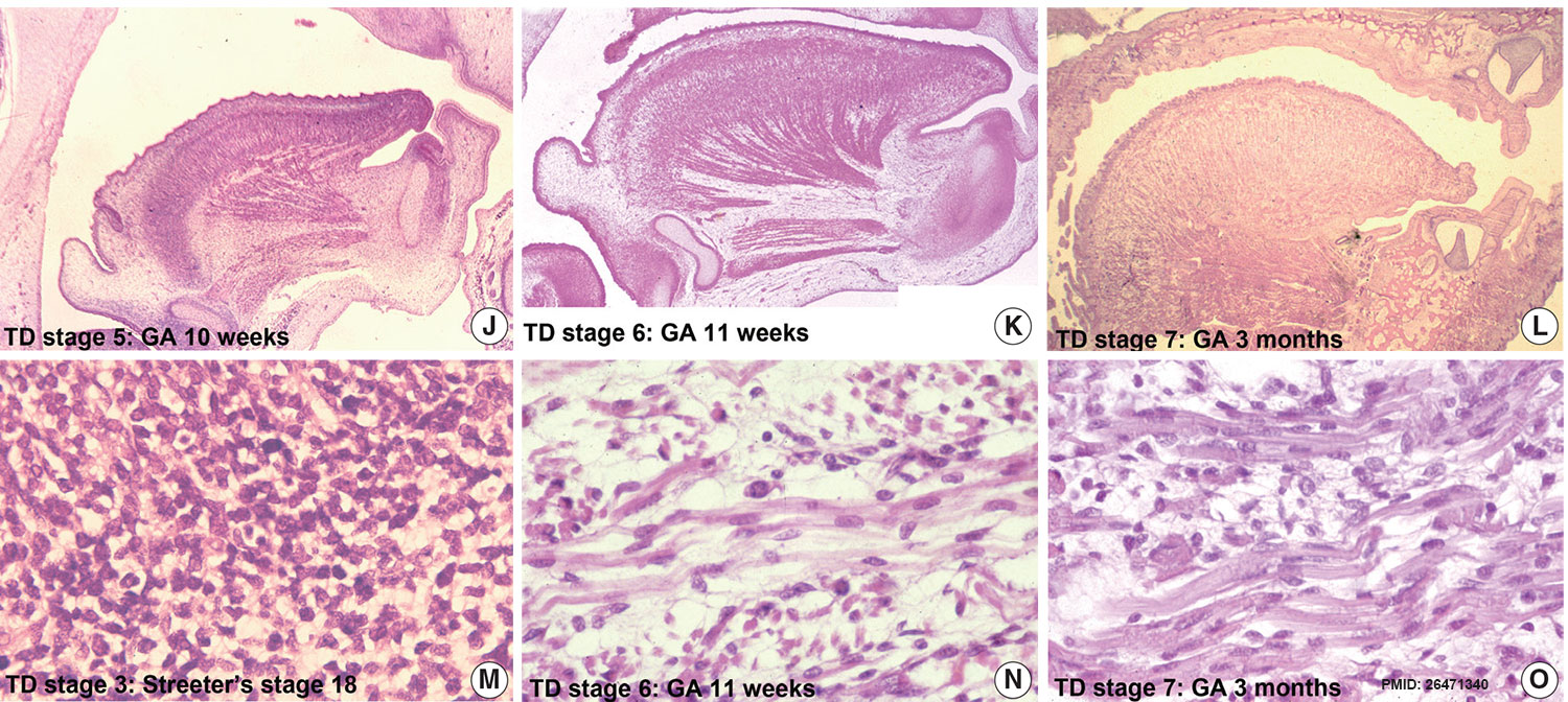
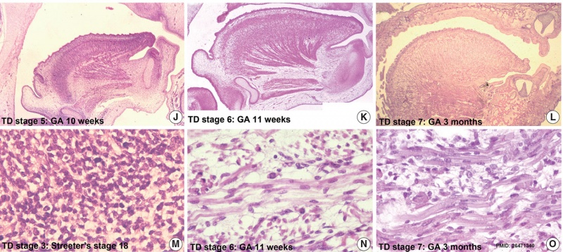
Organogenesis of a Fetal Tongue
Photographs of mid-sagittal sections of a fetal human tongue.
(I, J) TD stage 5. MC, Meckerl's cartilage; Md, mandible.
(K) TD stage 6.
(L) TD stage 7. Microscopic features of tongue muscle at TD stage 3 (M), TD stage 6 (N), and TD stage 7 (O).
- Tongue Links: image - embryonic | image - stage 13 (low and high) | image - stage 13 (low) | image - stage 13 (high) | image - stage 14 | image - stage 16 | image - stage 17 | image - stage 18 | image - stage 19 | image - stage 21 | image - stage 23 | image - fetal | image - embryo and fetal | Tongue Development | Taste Development | PMID26471340
Reference
Hong SJ, Cha BG, Kim YS, Lee SK & Chi JG. (2015). Tongue Growth during Prenatal Development in Korean Fetuses and Embryos. J Pathol Transl Med , 49, 497-510. PMID: 26471340 DOI.
Copyright
© 2015 The Korean Society of Pathologists/The Korean Society for Cytopathology This is an Open Access article distributed under the terms of the Creative Commons Attribution Non-Commercial License (http://creativecommons.org/licenses/by-nc/3.0/) which permits unrestricted non-commercial use, distribution, and reproduction in any medium, provided the original work is properly cited.
Fig. 1. Jptm-2015-09-17f1.jpg
Cite this page: Hill, M.A. (2024, April 27) Embryology Human fetal tongue 01.jpg. Retrieved from https://embryology.med.unsw.edu.au/embryology/index.php/File:Human_fetal_tongue_01.jpg
- © Dr Mark Hill 2024, UNSW Embryology ISBN: 978 0 7334 2609 4 - UNSW CRICOS Provider Code No. 00098G
File history
Click on a date/time to view the file as it appeared at that time.
| Date/Time | Thumbnail | Dimensions | User | Comment | |
|---|---|---|---|---|---|
| current | 13:36, 23 March 2016 | | 1,500 × 672 (350 KB) | Z8600021 (talk | contribs) |
You cannot overwrite this file.
File usage
The following page uses this file: