File:Human extraocular muscles 01.jpg
Human_extraocular_muscles_01.jpg (500 × 600 pixels, file size: 47 KB, MIME type: image/jpeg)
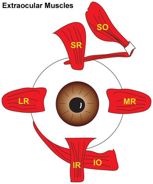
Human Extraocular Muscles
Cartoon showing attachment of the human 6 extraocular muscles to the eyeball.
| Legend | About the Muscles |
|---|---|
|
|
| Muscle Structure | ||
|---|---|---|
| Each muscle has 2 equally sized layers. | ||
| Layer | Inserts | Single innervation (%) |
| outer orbital | connective tissue (pulley) ring | 80 |
| inner global | eye sclera | 90 |
Reference
<pubmed>22132088</pubmed>| PLoS One.
Citation: Kasprick DS, Kish PE, Junttila TL, Ward LA, Bohnsack BL, et al. (2011) Microanatomy of Adult Zebrafish Extraocular Muscles. PLoS ONE 6(11): e27095. doi:10.1371/journal.pone.0027095
Copyright
© 2011 Kasprick et al. This is an open-access article distributed under the terms of the Creative Commons Attribution License, which permits unrestricted use, distribution, and reproduction in any medium, provided the original author and source are credited.
Figure 1. doi:10.1371/journal.pone.0027095.g001 Pone.0027095.g001.jpg Text modified from figure legend and paper text.
Cite this page: Hill, M.A. (2024, April 26) Embryology Human extraocular muscles 01.jpg. Retrieved from https://embryology.med.unsw.edu.au/embryology/index.php/File:Human_extraocular_muscles_01.jpg
- © Dr Mark Hill 2024, UNSW Embryology ISBN: 978 0 7334 2609 4 - UNSW CRICOS Provider Code No. 00098G
File history
Click on a date/time to view the file as it appeared at that time.
| Date/Time | Thumbnail | Dimensions | User | Comment | |
|---|---|---|---|---|---|
| current | 12:04, 8 June 2012 | | 500 × 600 (47 KB) | Z8600021 (talk | contribs) | ==Human Extraocular Muscles== Illustration of human eye showing 6 EOMs inserting on the globe in what is referred to as the Spiral of Tillaux. ===Reference== <pubmed>22132088</pubmed>| [http://www.plosone.org/article/info%3Adoi%2F10.1371%2Fjournal.pone |
You cannot overwrite this file.
File usage
The following 4 pages use this file: