File:Human embryo skin 8-9 week EGA.jpg: Difference between revisions
No edit summary |
m (→Reference) |
||
| (2 intermediate revisions by one other user not shown) | |||
| Line 1: | Line 1: | ||
==Human Embryo Skin (8-9 week EGA)== | ==Human Embryo Skin (8-9 week EGA)== | ||
TEM of embryonic epidermis showing periderm and basal layer. | Transmission electron micrograph (TEM) of embryonic epidermis showing periderm and basal layer. | ||
Desmosomes are circled and enlarged in k, and the area of the basal cell enlarged in i is enclosed within the box. Note that cells of both layers are filled primarily with glycogen. (I) TEM of a periderm cell showing fine bundles of keratin filaments (arrows). | Desmosomes are circled and [[:File:Human_embryo_skin_8-9_week_EGA_desmosomes.jpg|enlarged in k]], and the area of the basal cell enlarged in i is enclosed within the box. Note that cells of both layers are filled primarily with glycogen. (I) TEM of a periderm cell showing fine bundles of keratin filaments (arrows). | ||
* 8 to 9 weeks Estimated Gestational Age = about 6 to 8 weeks Fertilization Age | * 8 to 9 weeks Estimated Gestational Age = about 6 to 8 weeks Fertilization Age | ||
| Line 10: | Line 10: | ||
{{Epidermis EM links}} | {{Epidermis EM links}} | ||
===Reference=== | ===Reference=== | ||
{{#pmid:2413039}} | |||
{{JCB}} | {{JCB}} | ||
{{Footer}} | |||
[[Category:Integumentary]] | |||
Latest revision as of 12:23, 27 March 2019
Human Embryo Skin (8-9 week EGA)
Transmission electron micrograph (TEM) of embryonic epidermis showing periderm and basal layer.
Desmosomes are circled and enlarged in k, and the area of the basal cell enlarged in i is enclosed within the box. Note that cells of both layers are filled primarily with glycogen. (I) TEM of a periderm cell showing fine bundles of keratin filaments (arrows).
- 8 to 9 weeks Estimated Gestational Age = about 6 to 8 weeks Fertilization Age
- Magnification x 37,000 (image cropped from figure 1, contrast adjusted)
- Links: Epidermis 8-9 week EGA | Epidermis desmosomes 8-9 week EGA | Epidermis 9-11 week EGA | Epidermis 24 week EGA | Integumentary System Development
Reference
Dale BA, Holbrook KA, Kimball JR, Hoff M & Sun TT. (1985). Expression of epidermal keratins and filaggrin during human fetal skin development. J. Cell Biol. , 101, 1257-69. PMID: 2413039
Copyright
Rockefeller University Press - Copyright Policy This article is distributed under the terms of an Attribution–Noncommercial–Share Alike–No Mirror Sites license for the first six months after the publication date (see http://www.jcb.org/misc/terms.shtml). After six months it is available under a Creative Commons License (Attribution–Noncommercial–Share Alike 4.0 Unported license, as described at https://creativecommons.org/licenses/by-nc-sa/4.0/ ). (More? Help:Copyright Tutorial)
Cite this page: Hill, M.A. (2024, May 5) Embryology Human embryo skin 8-9 week EGA.jpg. Retrieved from https://embryology.med.unsw.edu.au/embryology/index.php/File:Human_embryo_skin_8-9_week_EGA.jpg
- © Dr Mark Hill 2024, UNSW Embryology ISBN: 978 0 7334 2609 4 - UNSW CRICOS Provider Code No. 00098G
File history
Click on a date/time to view the file as it appeared at that time.
| Date/Time | Thumbnail | Dimensions | User | Comment | |
|---|---|---|---|---|---|
| current | 22:49, 28 September 2011 | 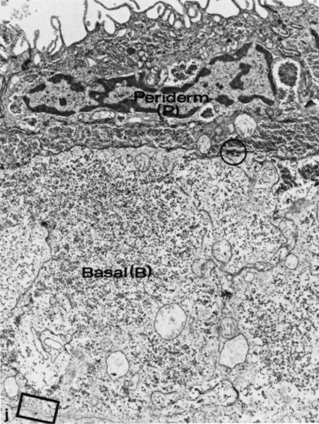 | 657 × 872 (188 KB) | S8600021 (talk | contribs) | ==Human Embryo Skin (8-9 week EGA)== TEM of embryonic epidermis showing periderm and basal layer. Desmosomes are circled and enlarged in k, and the area of the basal cell enlarged in i is enclosed within the box. Note that cells of both layers are fill |
You cannot overwrite this file.
File usage
The following 4 pages use this file: