File:Human congenital diaphragmatic hernia.jpg

Original file (800 × 626 pixels, file size: 86 KB, MIME type: image/jpeg)
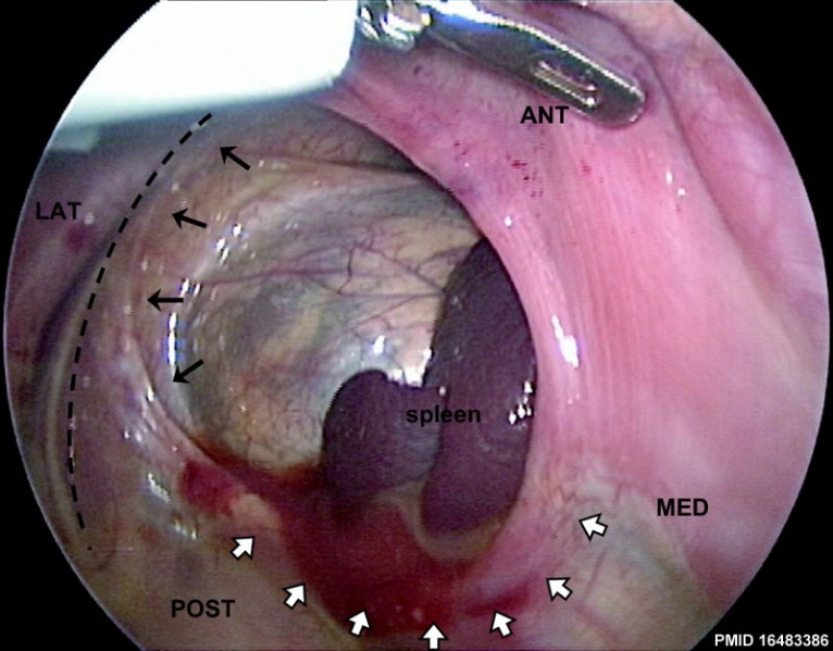
Human Congenital Diaphragmatic Hernia (CDH)
View of a human CDH during thoracoscopic surgical repair. Commonly failure of the pleuroperitoneal foramen (foramen of Bochdalek) to close on the left side.
The image is obtained through the scope, from the chest (i.e. above) and with the infant rotated on the operating table. Hence, the image is slightly rotated and left-right reversed with respect to other figures within the original article. The retroperitoneum and spleen are visualized through the defect.
|
|
Surgical closure of the defect involves apposing diaphragm to chest wall laterally but diaphragm to diaphragm medially. Larger defects may not be amenable to primary closure and generally are repaired with a prosthetic patch.
Reference
Fisher JC & Bodenstein L. (2006). Computer simulation analysis of normal and abnormal development of the mammalian diaphragm. Theor Biol Med Model , 3, 9. PMID: 16483386 DOI.
Fisher and Bodenstein Theoretical Biology and Medical Modelling 2006 3:9 doi:10.1186/1742-4682-3-9
Copyright
© 2006 Fisher and Bodenstein; licensee BioMed Central Ltd.
This is an Open Access article distributed under the terms of the Creative Commons Attribution License (http://creativecommons.org/licenses/by/2.0), which permits unrestricted use, distribution, and reproduction in any medium, provided the original work is properly cited.
Image courtesy of Dr. Edmund Yang, Vanderbilt Children's Hospital, Nashville, TN, USA., text modified from figure legend. 1742-4682-3-9-2-l.jpg
Cite this page: Hill, M.A. (2024, April 27) Embryology Human congenital diaphragmatic hernia.jpg. Retrieved from https://embryology.med.unsw.edu.au/embryology/index.php/File:Human_congenital_diaphragmatic_hernia.jpg
- © Dr Mark Hill 2024, UNSW Embryology ISBN: 978 0 7334 2609 4 - UNSW CRICOS Provider Code No. 00098G
File history
Click on a date/time to view the file as it appeared at that time.
| Date/Time | Thumbnail | Dimensions | User | Comment | |
|---|---|---|---|---|---|
| current | 18:27, 9 July 2012 |  | 800 × 626 (86 KB) | Z8600021 (talk | contribs) | ==Human Congenital Diaphragmatic Hernia== View of a human CDH during thoracoscopic surgical repair. The image is obtained through the scope, from the chest (i.e. above) and with the infant rotated on the operating table. Hence, the image is slightly rota |
You cannot overwrite this file.
File usage
The following 5 pages use this file: