File:Human blastocyst derived stem cells.jpg
From Embryology
Size of this preview: 800 × 550 pixels. Other resolution: 1,200 × 825 pixels.
Original file (1,200 × 825 pixels, file size: 198 KB, MIME type: image/jpeg)
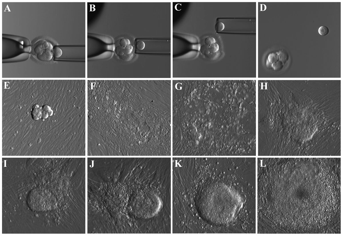
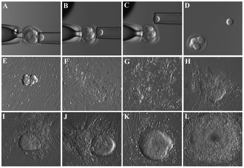
Derivation of Human Embryonic Stem Cells (hESC) from biopsied blastomere of the cleavage stage embryo
- A to D - micrographs showing the stepwise procedure of embryo biopsy using inverted microscope-attached micro manipulator.
- E to L - appearance of initial outgrowth and hESC colony during the derivation procedure.
All pictures were taken using microscope mounted digital camera and Hoffman optics under 200X total magnification.
- Links: Blastocyst | Stem Cells
Reference
<pubmed>22039509</pubmed>| PLoS One.
Copyright: © 2011 Giritharan et al. This is an open-access article distributed under the terms of the Creative Commons Attribution License, which permits unrestricted use, distribution, and reproduction in any medium, provided the original author and source are credited.
Figure 1. doi:10.1371/journal.pone.0026570.g001
File history
Click on a date/time to view the file as it appeared at that time.
| Date/Time | Thumbnail | Dimensions | User | Comment | |
|---|---|---|---|---|---|
| current | 21:50, 7 November 2011 | | 1,200 × 825 (198 KB) | S8600021 (talk | contribs) | == Derivation of Human Embryonic Stem Cells (hESC) from biopsied blastomere of the cleavage stage embryo== Micrographs showing the stepwise procedure of embryo biopsy using inverted microscope-attached micromanipulator (A–D) and the appearance of initi |
You cannot overwrite this file.
File usage
The following page uses this file: