File:Human-spermatozoa EM01.jpg: Difference between revisions
No edit summary |
mNo edit summary |
||
| Line 1: | Line 1: | ||
==Human Spermatozoa ( transmission electron micrograph)== | |||
Montage transmission electron micrograph of a human sperm cell. | Montage transmission electron micrograph of a human sperm cell. | ||
The cell has a compact nucleus, conspicuous mitochondria, no endoplasmic reticulum, minimal cytoplasm and a large tail (about 45 μm in length). Superfluous cytoplasm and associated machinery is jettisoned when the sperm emerges from the testis, leaving a 'stripped down', minimalist cell. | The cell has a compact nucleus, conspicuous mitochondria, no endoplasmic reticulum, minimal cytoplasm and a large tail (about 45 μm in length). Superfluous cytoplasm and associated machinery is jettisoned when the sperm emerges from the testis, leaving a 'stripped down', minimalist cell. | ||
===Reference=== | |||
<pubmed>19678911</pubmed>| [http://www.ncbi.nlm.nih.gov/pmc/articles/PMC2736672/ PMC: 2736672]] | [http://jbiol.com/content/8/7/63 J of Biology] | |||
{{Template:BMC}} | {{Template:BMC}} | ||
Barratt et al. Journal of Biology 2009 8:63 doi:10.1186/jbiol167 | |||
[[Category:Spermatozoa]] [[Category:Fertilization]] [[Category:Electron Micrograph]] [[Category:Male]] | [[Category:Spermatozoa]] [[Category:Fertilization]] [[Category:Electron Micrograph]] [[Category:Male]] | ||
Revision as of 13:28, 20 April 2013
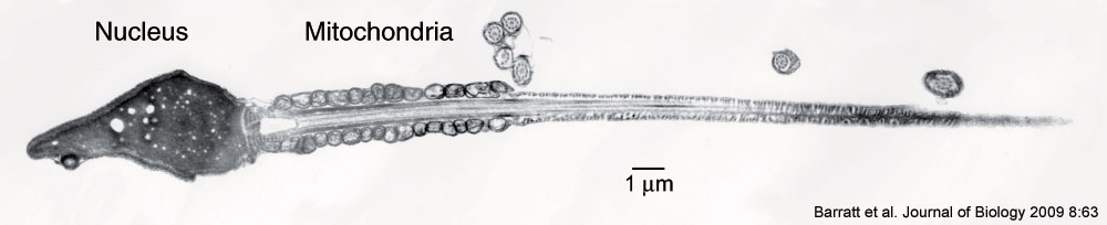
Human Spermatozoa ( transmission electron micrograph)
Montage transmission electron micrograph of a human sperm cell.
The cell has a compact nucleus, conspicuous mitochondria, no endoplasmic reticulum, minimal cytoplasm and a large tail (about 45 μm in length). Superfluous cytoplasm and associated machinery is jettisoned when the sperm emerges from the testis, leaving a 'stripped down', minimalist cell.
Reference
<pubmed>19678911</pubmed>| PMC: 2736672] | J of Biology
BioMed Central Open Access BioMed Central Open Access license agreement Brief summary of the agreement.
Anyone is free: to copy, distribute, and display the work; to make derivative works; to make commercial use of the work.
Under the following conditions: Attribution, the original author must be given credit; for any reuse or distribution, it must be made clear to others what the license terms of this work are; any of these conditions can be waived if the authors gives permission.
Barratt et al. Journal of Biology 2009 8:63 doi:10.1186/jbiol167
File history
Click on a date/time to view the file as it appeared at that time.
| Date/Time | Thumbnail | Dimensions | User | Comment | |
|---|---|---|---|---|---|
| current | 13:24, 5 April 2010 | 1,000 × 204 (26 KB) | S8600021 (talk | contribs) | Montage transmission electron micrograph of a human sperm cell. The cell has a compact nucleus, conspicuous mitochondria, no endoplasmic reticulum, minimal cytoplasm and a large tail (about 45 μm in length). Superfluous cytoplasm and associated machine |
You cannot overwrite this file.