File:Human-retina-01.jpg
Original file (1,000 × 615 pixels, file size: 197 KB, MIME type: image/jpeg)
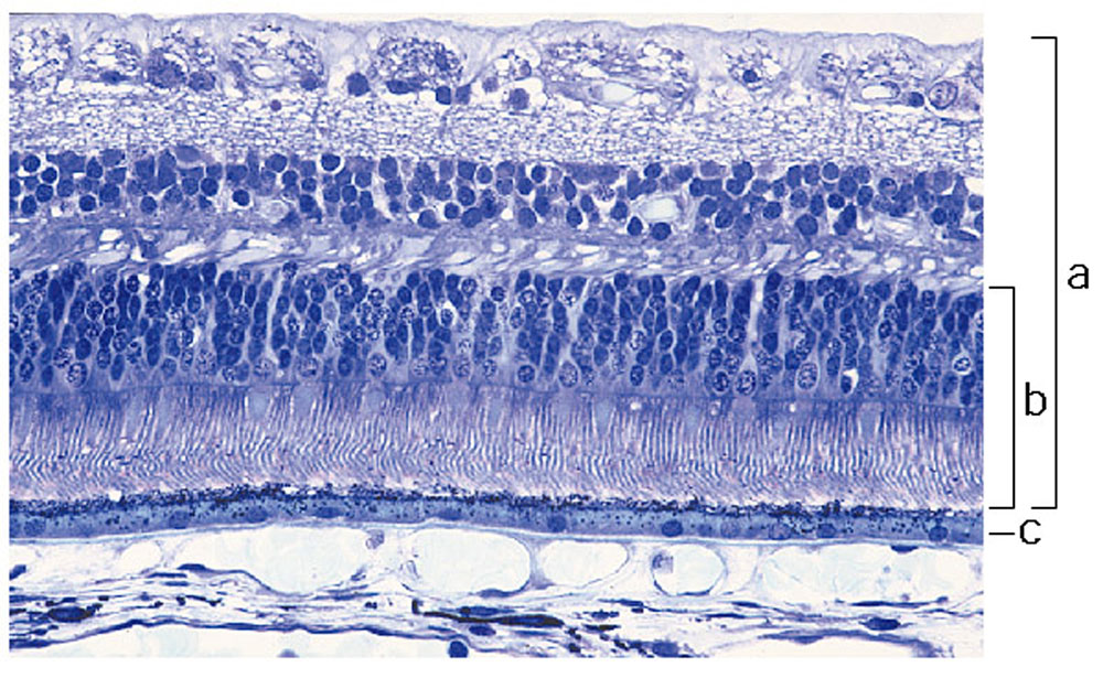
Adult Human Retina Histology
Light micrograph of normal human ((retina}} stained with Richardson's methylene blue/azure II. Light enters the retina from the top of the image.
(a) Neural retina
(b) photoreceptor layer
(c) retinal pigment epithelium (RPE)
Reference
{{#pmid12186651}}
From the Human Retina Teaching Set, Scheie Eye Institute, University of Pennsylvania, Philadelphia, USA. Courtesy of Ann Milam.
Copyright
Original Image Name: Figure 1. http://genomebiology.com/2002/3/8/reviews/1022/figure/F1
Cite this page: Hill, M.A. (2024, April 27) Embryology Human-retina-01.jpg. Retrieved from https://embryology.med.unsw.edu.au/embryology/index.php/File:Human-retina-01.jpg
- © Dr Mark Hill 2024, UNSW Embryology ISBN: 978 0 7334 2609 4 - UNSW CRICOS Provider Code No. 00098G
File history
Click on a date/time to view the file as it appeared at that time.
| Date/Time | Thumbnail | Dimensions | User | Comment | |
|---|---|---|---|---|---|
| current | 14:56, 16 October 2010 | | 1,000 × 615 (197 KB) | S8600021 (talk | contribs) | ==Adult Human Retina Histology== Light micrograph of normal human retina stained with Richardson's methylene blue/azure II. Light enters the retina from the top of the image. (a) Neural retina (b) photoreceptor layer (c) retinal pigment epithelium (RP |
You cannot overwrite this file.
File usage
The following 2 pages use this file: