File:Human- neural Chiari malformation.jpg
Original file (1,200 × 1,344 pixels, file size: 212 KB, MIME type: image/jpeg)
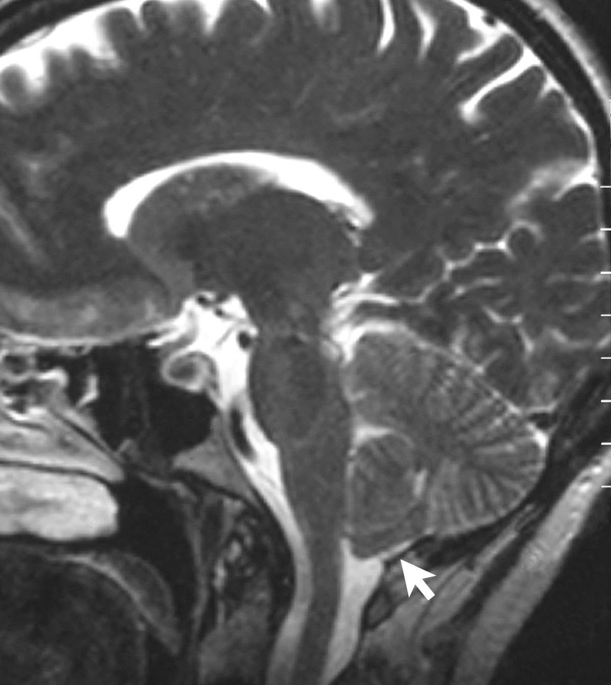
Chiari I malformation (CMI)
A T2-weighted sagittal MRI scan, from a patient with Chiari-like symptomatology, demonstrating tonsillar herniation less than 3 mm. The cisterna magna is completely obliterated by the thickened posterior rim of the foramen magnum. Meniscus sign (white arrow) is present at the inferior pole of the cerebellar tonsils consistent with a CSF block.
Original File Name: 1743-8454-2-11-1-l.jpg
Dimensions of the posterior fossa in patients symptomatic for Chiari I malformation but without cerebellar tonsillar descent. Sekula RF Jr, Jannetta PJ, Casey KF, Marchan EM, Sekula LK, McCrady CS. Cerebrospinal Fluid Res. 2005 Dec 18;2:11. PMID: 16359556 | Cerebrospinal Fluid Res.
© 2005 Sekula et al; licensee BioMed Central Ltd.
This is an Open Access article distributed under the terms of the Creative Commons Attribution License (http://creativecommons.org/licenses/by/2.0), which permits unrestricted use, distribution, and reproduction in any medium, provided the original work is properly cited.
File history
Click on a date/time to view the file as it appeared at that time.
| Date/Time | Thumbnail | Dimensions | User | Comment | |
|---|---|---|---|---|---|
| current | 12:03, 28 April 2010 | | 1,200 × 1,344 (212 KB) | S8600021 (talk | contribs) | Chiari I malformation (CMI) A T2-weighted sagittal MRI scan, from a patient with Chiari-like symptomatology, demonstrating tonsillar herniation less than 3 mm. The cisterna magna is completely obliterated by the thickened posterior rim of the foramen mag |
You cannot overwrite this file.
File usage
There are no pages that use this file.