File:Human- adult brain MRI.jpg

Original file (1,200 × 1,170 pixels, file size: 160 KB, MIME type: image/jpeg)
Adult Human Brain MRI
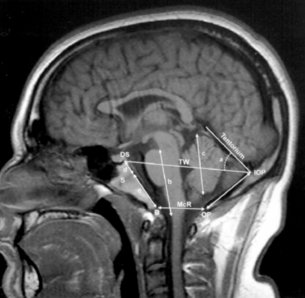
A T1-weighted sagittal MR image from a control subject, showing the midline structures of the posterior cranial fossa and the brainstem and the cerebellum.
- d + e = length of clivus
- S = sphenooccipital synchondrosis
- d = length of basisphenoid between the top of the dorsum sellae and the sphenooccipital synchondrosis of the clivus
- e = length of the basiocciput between the synchondrosis and the basion
- b = length of the hindbrain between the midbrain-pons junction and the medullocervical junction
- a = angle of the cerebellar tentorium to Twining's line
- c = length of cerebellar hemisphere
- DS = top of the dorsum sellae
- IOP = internal occipital protuberance
- OP = opisthion; IOP to OP = length of supraocciput
- B = basion; TW = Twining's line
- McR (B to OP) = McRae's line
Reference
<pubmed>16359556</pubmed>| Cerebrospinal Fluid Res.
Copyright
© 2005 Sekula et al; licensee BioMed Central Ltd.
This is an Open Access article distributed under the terms of the Creative Commons Attribution License (http://creativecommons.org/licenses/by/2.0), which permits unrestricted use, distribution, and reproduction in any medium, provided the original work is properly cited.
Original File Name: 1743-8454-2-11-2-l.jpg
File history
Click on a date/time to view the file as it appeared at that time.
| Date/Time | Thumbnail | Dimensions | User | Comment | |
|---|---|---|---|---|---|
| current | 12:26, 28 April 2010 | | 1,200 × 1,170 (160 KB) | S8600021 (talk | contribs) | Human- adult brain MRI A T1-weighted sagittal MR image from a control subject, showing the midline structures of the posterior cranial fossa and the brainstem and the cerebellum. * d + e = length of clivus * S = sphenooccipital synchondrosis * d = leng |
You cannot overwrite this file.
File usage
The following page uses this file: