File:Hughes1950 fig03.jpg
From Embryology

Size of this preview: 396 × 599 pixels. Other resolution: 900 × 1,362 pixels.
Original file (900 × 1,362 pixels, file size: 194 KB, MIME type: image/jpeg)
Fig. 3. Human Embryo 9 to 10 mm CRL
A layer of mesoderm cells quickly condenses deep to the mammary crest. Deep to this again is a widening zone of condensed mesoderm.
Reference
Hughes ES. Development of the mammary gland. (1950) Ann R Coll Surg Engl. 6(2):99-119. PMID 19309885
Cite this page: Hill, M.A. (2024, April 27) Embryology Hughes1950 fig03.jpg. Retrieved from https://embryology.med.unsw.edu.au/embryology/index.php/File:Hughes1950_fig03.jpg
- © Dr Mark Hill 2024, UNSW Embryology ISBN: 978 0 7334 2609 4 - UNSW CRICOS Provider Code No. 00098G
File history
Click on a date/time to view the file as it appeared at that time.
| Date/Time | Thumbnail | Dimensions | User | Comment | |
|---|---|---|---|---|---|
| current | 10:18, 15 August 2018 | | 900 × 1,362 (194 KB) | Z8600021 (talk | contribs) | |
| 10:16, 15 August 2018 | 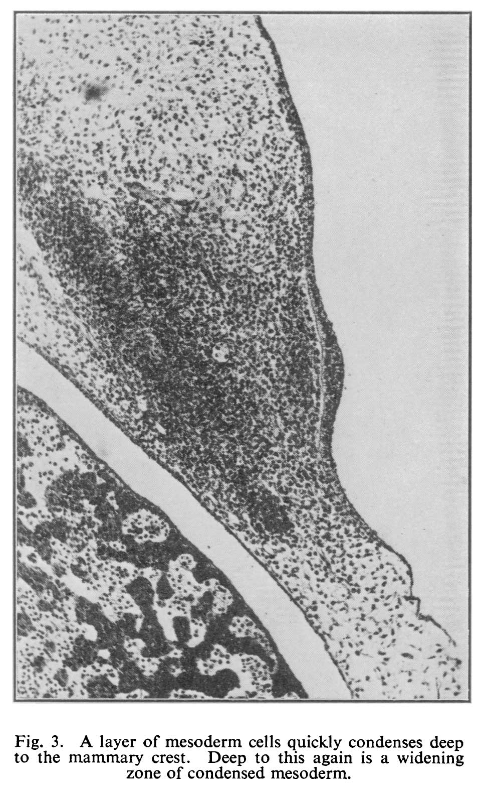 | 964 × 1,576 (215 KB) | Z8600021 (talk | contribs) | Fig. 3. A layer of mesoderm cells quickly condenses deep to the mammary crest. Deep to this again is a widening zone of condensed mesoderm. ===Reference=== {{Ref-Hughes1950}} {{Footer}} |
You cannot overwrite this file.
File usage
The following page uses this file: