File:Horseshoe.jpg
From Embryology
No higher resolution available.
Horseshoe.jpg (400 × 400 pixels, file size: 32 KB, MIME type: image/jpeg)
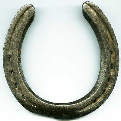
A Horseshoe
The term "horseshoe" is used to describe the renal abnormality horseshoe kidney where there is typically a fusion of the lower poles of both kidneys. This gives the fused structure the shape of a horseshoe.
I also have used this term to describe the shape of the [[? intra-embryonic coelom within the lateral plate mesoderm that forms during week 3 of human development.
Cite this page: Hill, M.A. (2024, April 26) Embryology Horseshoe.jpg. Retrieved from https://embryology.med.unsw.edu.au/embryology/index.php/File:Horseshoe.jpg
- © Dr Mark Hill 2024, UNSW Embryology ISBN: 978 0 7334 2609 4 - UNSW CRICOS Provider Code No. 00098G
File history
Click on a date/time to view the file as it appeared at that time.
| Date/Time | Thumbnail | Dimensions | User | Comment | |
|---|---|---|---|---|---|
| current | 12:34, 15 September 2012 | | 400 × 400 (32 KB) | Z8600021 (talk | contribs) | ==A Horseshoe== The horseshoe is used to describe the renal abnormality horseshoe kidney there is typically a fusion of the lower poles of both kidneys. :'''Links:''' [[Renal_System_-_Abnormalities|Renal Abnormalitie |
You cannot overwrite this file.
File usage
The following page uses this file: