File:Heart histology 001.jpg: Difference between revisions
No edit summary |
|||
| (3 intermediate revisions by the same user not shown) | |||
| Line 1: | Line 1: | ||
==Heart Histology== | ==Heart Histology - Purkinje Fibre== | ||
* sheep cardiac muscle, transverse section | * sheep cardiac muscle, transverse section | ||
* Purkinje Fibre | * Purkinje Fibre. | ||
* Stain: Whipf's polychrome, x40 | * Stain: Whipf's polychrome, x40 | ||
===Purkinje Fibres=== | |||
* modified cardiac muscle cells. Compared to ordinary cardiac muscle cells: | |||
** contain large amounts of glycogen. | |||
** fewer myofibrils. | |||
** thicker cells. | |||
* extend from the '''atrioventricular node''', pierces the fibrous body, divides into left and right bundles, and travels, beneath the endocardium, towards the apex of the heart. | |||
* bundle branches contact cardiac muscle cells through specialisations similar to intercalated discs. | |||
* conduct stimuli faster than ordinary cardiac muscle cells (2-3 m/s vs. 0.6 m/s). | |||
* discovered in 1839 by Jan Evangelista Purkyně). | |||
Latest revision as of 08:11, 6 August 2012
Heart Histology - Purkinje Fibre
- sheep cardiac muscle, transverse section
- Purkinje Fibre.
- Stain: Whipf's polychrome, x40
Purkinje Fibres
- modified cardiac muscle cells. Compared to ordinary cardiac muscle cells:
- contain large amounts of glycogen.
- fewer myofibrils.
- thicker cells.
- extend from the atrioventricular node, pierces the fibrous body, divides into left and right bundles, and travels, beneath the endocardium, towards the apex of the heart.
- bundle branches contact cardiac muscle cells through specialisations similar to intercalated discs.
- conduct stimuli faster than ordinary cardiac muscle cells (2-3 m/s vs. 0.6 m/s).
- discovered in 1839 by Jan Evangelista Purkyně).
Whipf's polychrome staining procedure - used by Dr. Louise Whipf, Veterinary Pathology Laboratory, University of Wisconsin, who originally modified Shorr’s (1941) stain.
- Links: Heart Histology | Cardiac AZB Labeled | Cardiac AZB | Cardiac label LS | Cardiac LS | Cardiac label TS | Cardiac TS | Purkinje fibres | Purkinje fibres detail | Histology
Links: Histology | Histology Stains | Blue Histology images copyright Lutz Slomianka 1998-2009. The literary and artistic works on the original Blue Histology website may be reproduced, adapted, published and distributed for non-commercial purposes. See also the page Histology Stains.
Cite this page: Hill, M.A. (2024, May 1) Embryology Heart histology 001.jpg. Retrieved from https://embryology.med.unsw.edu.au/embryology/index.php/File:Heart_histology_001.jpg
- © Dr Mark Hill 2024, UNSW Embryology ISBN: 978 0 7334 2609 4 - UNSW CRICOS Provider Code No. 00098G
Original file name: Cardm042wp.jpg
Cite this page: Hill, M.A. (2024, May 1) Embryology Heart histology 001.jpg. Retrieved from https://embryology.med.unsw.edu.au/embryology/index.php/File:Heart_histology_001.jpg
- © Dr Mark Hill 2024, UNSW Embryology ISBN: 978 0 7334 2609 4 - UNSW CRICOS Provider Code No. 00098G
File history
Click on a date/time to view the file as it appeared at that time.
| Date/Time | Thumbnail | Dimensions | User | Comment | |
|---|---|---|---|---|---|
| current | 09:25, 14 August 2011 | 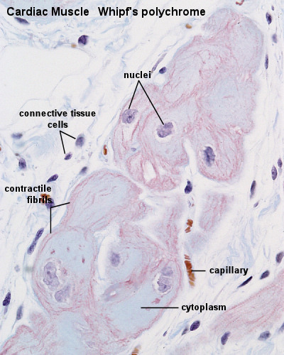 | 400 × 500 (83 KB) | S8600021 (talk | contribs) | ==Heart Histology== cardiac muscle, Purkinje Fibre, sheep - Whipf's polychrome transverse section, x40 Original file name: Cardm042wp.jpg {{Blue Histology}} {{Footer}} Category:Human |
You cannot overwrite this file.
File usage
The following 6 pages use this file: