File:Hair follicle cell development.png
Original file (2,012 × 681 pixels, file size: 1.44 MB, MIME type: image/png)
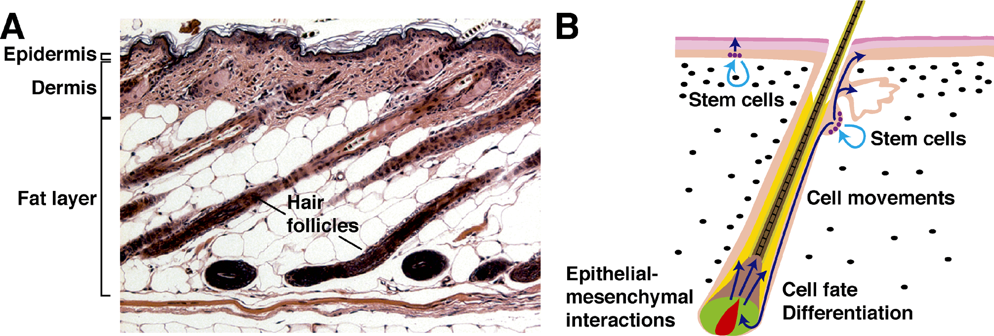
(A) Hematoxylin/eosin-stained section of mouse skin at postnatal day 28, showing hair follicles in the anagen growth phase; major layers of the skin are indicated.
(B) Schematic depiction of postnatal skin showing a hair follicle in the growth phase. Biological processes occurring in the skin are listed and SC locations are indicated. Dark blue arrows indicate the movements of stem and matrix cell progeny; pale blue arrows indicate SC self-renewal. Pink, epidermis and hair follicle outer root sheath; yellow, inner root sheath; green, matrix; red, hair follicle DP; light brown, hair shaft precursors; darker brown, hair shaft; violet circles, SCs; black ovals, dermal fibroblasts.
http://www.plosbiology.org/article/info:doi/10.1371/journal.pbio.0030372
Citation: Millar SE (2005) An Ideal Society? Neighbors of Diverse Origins Interact to Create and Maintain Complex Mini-Organs in the Skin. PLoS Biol 3(11): e372. doi:10.1371/journal.pbio.0030372
Published: November 15, 2005
Copyright: © 2005 Sarah E. Millar. This is an open-access article distributed under the terms of the Creative Commons Attribution License, which permits unrestricted use, distribution, and reproduction in any medium, provided the original author and source are credited.
File history
Click on a date/time to view the file as it appeared at that time.
| Date/Time | Thumbnail | Dimensions | User | Comment | |
|---|---|---|---|---|---|
| current | 08:14, 29 September 2009 | 2,012 × 681 (1.44 MB) | S8600021 (talk | contribs) | (A) Hematoxylin/eosin-stained section of mouse skin at postnatal day 28, showing hair follicles in the anagen growth phase; major layers of the skin are indicated. (B) Schematic depiction of postnatal skin showing a hair follicle in the growth phase. Bio |
You cannot overwrite this file.
File usage
The following 2 pages use this file: