File:Gray1016.jpg
Gray1016.jpg (252 × 400 pixels, file size: 18 KB, MIME type: image/jpeg)
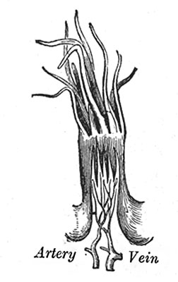
Fig. 1016. A Filiform Papilla
The papillæ filiformes (filiform or conical papilæ) (Fig. 1016) cover the anterior two-thirds of the dorsum. They are very minute, filiform in shape, and arranged in lines parallel with the two rows of the papillæ vallatæ, excepting at the apex of the organ, where their direction is transverse. Projecting from their apices are numerous filamentous processes, or secondary papillæ these are of a whitish tint, owing to the thickness and density of the epithelium of which they are composed, which has here undergone a peculiar modification, the cells having become cornified and elongated into dense, imbricated, brush-like processes. They contain also a number of elastic fibers, which render them firmer and more elastic than the papillæ of mucous membrane generally. The larger and longer papillæ of this group are sometimes termed papilla conicæ.
- Links: Tongue Development | Taste Development | Head Development |
- Gray's Images: Development | Lymphatic | Neural | Vision | Hearing | Somatosensory | Integumentary | Respiratory | Gastrointestinal | Urogenital | Endocrine | Surface Anatomy | iBook | Historic Disclaimer
| Historic Disclaimer - information about historic embryology pages |
|---|
| Pages where the terms "Historic" (textbooks, papers, people, recommendations) appear on this site, and sections within pages where this disclaimer appears, indicate that the content and scientific understanding are specific to the time of publication. This means that while some scientific descriptions are still accurate, the terminology and interpretation of the developmental mechanisms reflect the understanding at the time of original publication and those of the preceding periods, these terms, interpretations and recommendations may not reflect our current scientific understanding. (More? Embryology History | Historic Embryology Papers) |
| iBook - Gray's Embryology | |
|---|---|

|
|
Reference
Gray H. Anatomy of the human body. (1918) Philadelphia: Lea & Febiger.
Cite this page: Hill, M.A. (2024, April 27) Embryology Gray1016.jpg. Retrieved from https://embryology.med.unsw.edu.au/embryology/index.php/File:Gray1016.jpg
- © Dr Mark Hill 2024, UNSW Embryology ISBN: 978 0 7334 2609 4 - UNSW CRICOS Provider Code No. 00098G
File history
Click on a date/time to view the file as it appeared at that time.
| Date/Time | Thumbnail | Dimensions | User | Comment | |
|---|---|---|---|---|---|
| current | 08:13, 11 May 2014 |  | 252 × 400 (18 KB) | Z8600021 (talk | contribs) | :'''Links:''' Tongue Development | Taste Development | Head Development | {{Gray Anatomy}} Category:Tongue Category:Head Category:Cartoon |
You cannot overwrite this file.
File usage
The following 2 pages use this file:

