File:Gray0969.jpg

Original file (621 × 900 pixels, file size: 241 KB, MIME type: image/jpeg)
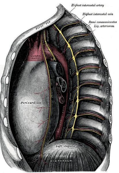
Middle and Posterior Mediastina
View Left side.
The Middle Mediastinum (Fig. 968) is the broadest part of the interpleural space. It contains the heart enclosed in the pericardium, the ascending aorta, the lower half of the superior vena cava with the azygos vein opening into it, the bifurcation of the trachea and the two bronchi, the pulmonary artery dividing into its two branches, the right and left pulmonary veins, the phrenic nerves, and some bronchial lymph glands.
The Posterior Mediastinum (Figs. 968, 969) is an irregular triangular space running parallel with the vertebral column; it is bounded in front by the pericardium above, and by the posterior surface of the diaphragm below, behind by the vertebral column from the lower border of the fourth to the twelfth thoracic vertebra, and on either side by the mediastinal pleura. It contains the thoracic part of the descending aorta, the azygos and the two hemiazygos veins, the vagus and splanchnic nerves, the esophagus, the thoracic duct, and some lymph glands.
- Gray's Images: Development | Lymphatic | Neural | Vision | Hearing | Somatosensory | Integumentary | Respiratory | Gastrointestinal | Urogenital | Endocrine | Surface Anatomy | iBook | Historic Disclaimer
| Historic Disclaimer - information about historic embryology pages |
|---|
| Pages where the terms "Historic" (textbooks, papers, people, recommendations) appear on this site, and sections within pages where this disclaimer appears, indicate that the content and scientific understanding are specific to the time of publication. This means that while some scientific descriptions are still accurate, the terminology and interpretation of the developmental mechanisms reflect the understanding at the time of original publication and those of the preceding periods, these terms, interpretations and recommendations may not reflect our current scientific understanding. (More? Embryology History | Historic Embryology Papers) |
| iBook - Gray's Embryology | |
|---|---|

|
|
Reference
Gray H. Anatomy of the human body. (1918) Philadelphia: Lea & Febiger.
Cite this page: Hill, M.A. (2024, April 27) Embryology Gray0969.jpg. Retrieved from https://embryology.med.unsw.edu.au/embryology/index.php/File:Gray0969.jpg
- © Dr Mark Hill 2024, UNSW Embryology ISBN: 978 0 7334 2609 4 - UNSW CRICOS Provider Code No. 00098G
File history
Click on a date/time to view the file as it appeared at that time.
| Date/Time | Thumbnail | Dimensions | User | Comment | |
|---|---|---|---|---|---|
| current | 03:38, 17 August 2012 |  | 621 × 900 (241 KB) | Z8600021 (talk | contribs) | ==Middle and Posterior Mediastina== View Left side. The Middle Mediastinum (Fig. 968) is the broadest part of the interpleural space. It contains the heart enclosed in the pericardium, the ascending aorta, the lower half of the superior vena cava with t |
You cannot overwrite this file.
File usage
The following 4 pages use this file:
