File:Gray0962.jpg

Original file (801 × 669 pixels, file size: 191 KB, MIME type: image/jpeg)
Bronchi and bronchioles
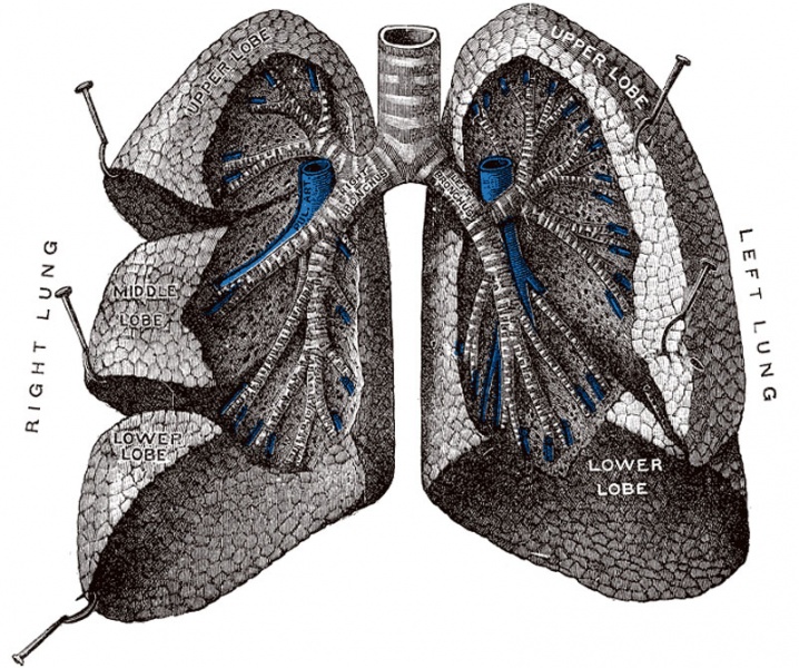
The lungs have been widely separated and tissue cut away to expose the air-tubes. (Testut.)
The trachea or windpipe (Fig. 961) is a cartilaginous and membranous tube, extending from the lower part of the larynx, on a level with the sixth cervical vertebra, to the upper border of the fifth thoracic vertebra, where it divides into the two bronchi, one for each lung. The trachea is nearly but not quite cylindrical, being flattened posteriorly; it measures about 11 cm. in length; its diameter, from side to side, is from 2 to 2.5 cm., being always greater in the male than in the female. In the child the trachea is smaller, more deeply placed, and more movable than in the adult.
Relations.—The anterior surface of the trachea is convex, and covered, in the neck, from above downward, by the isthmus of the thyroid gland, the inferior thyroid veins, the arteria thyroidea ima (when that vessel exists), the Sternothyreoideus and Sternohyoideus muscles, the cervical fascia, and, more superficially, by the anastomosing branches between the anterior jugular veins; in the thorax, it is covered from before backward by the manubrium sterni, the remains of the thymus, the left innominate vein, the aortic arch, the innominate and left common carotid arteries, and the deep cardiac plexus. Posteriorly it is in contact with the esophagus. Laterally, in the neck, it is in relation with the common carotid arteries, the right and left lobes of the thyroid gland, the inferior thyroid arteries, and the recurrent nerves; in the thorax, it lies in the superior mediastinum, and is in relation on the right side with the pleura and right vagus, and near the root of the neck with the innominate artery; on its left side are the left recurrent nerve, the aortic arch, and the left common carotid and subclavian arteries.
The Right Bronchus (bronchus dexter), wider, shorter, and more vertical in direction than the left, is about 2.5 cm. long, and enters the right lung nearly opposite the fifth thoracic vertebra. The azygos vein arches over it from behind; and the right pulmonary artery lies at first below and then in front of it. About 2 cm. from its commencement it gives off a branch to the upper lobe of the right lung. This is termed the eparterial branch of the bronchus, because it arises above the right pulmonary artery. The bronchus now passes below the artery, and is known as the hyparterial branch; it divides into two branches for the middle and lower lobes.
(Text modified from Gray's 1918 Anatomy)
- Larynx Image Links: All cartilages of the larynx | Epiglottis cartilage | Thyroid cartilage | Cricoid cartilage | Arytenoid cartilage | Larynx ligaments anterior | Larynx ligaments posterior | Larynx sagittal section | Larynx and upper trachea | Larynx entrance | Larynx interior | Larynx muscular attachments | Larynx muscles 1 | Larynx muscles 2 | Larynx muscles 3 | Cartilage Development | Respiratory System Development
- Gray's Images: Development | Lymphatic | Neural | Vision | Hearing | Somatosensory | Integumentary | Respiratory | Gastrointestinal | Urogenital | Endocrine | Surface Anatomy | iBook | Historic Disclaimer
| Historic Disclaimer - information about historic embryology pages |
|---|
| Pages where the terms "Historic" (textbooks, papers, people, recommendations) appear on this site, and sections within pages where this disclaimer appears, indicate that the content and scientific understanding are specific to the time of publication. This means that while some scientific descriptions are still accurate, the terminology and interpretation of the developmental mechanisms reflect the understanding at the time of original publication and those of the preceding periods, these terms, interpretations and recommendations may not reflect our current scientific understanding. (More? Embryology History | Historic Embryology Papers) |
| iBook - Gray's Embryology | |
|---|---|

|
|
Reference
Gray H. Anatomy of the human body. (1918) Philadelphia: Lea & Febiger.
Cite this page: Hill, M.A. (2024, April 27) Embryology Gray0962.jpg. Retrieved from https://embryology.med.unsw.edu.au/embryology/index.php/File:Gray0962.jpg
- © Dr Mark Hill 2024, UNSW Embryology ISBN: 978 0 7334 2609 4 - UNSW CRICOS Provider Code No. 00098G
File history
Click on a date/time to view the file as it appeared at that time.
| Date/Time | Thumbnail | Dimensions | User | Comment | |
|---|---|---|---|---|---|
| current | 03:51, 17 August 2012 |  | 801 × 669 (191 KB) | Z8600021 (talk | contribs) | {{Gray Anatomy}} |
You cannot overwrite this file.
File usage
The following 4 pages use this file:
