File:Gray0957.jpg

Original file (421 × 700 pixels, file size: 74 KB, MIME type: image/jpeg)
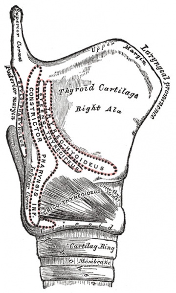
Larynx Muscular Attachments
Side view of Larynx.
The Cricothyreoideus (Cricothyroid) (Fig. 957), triangular in form, arises from the front and lateral part of the cricoid cartilage; its fibers diverge, and are arranged in two groups. The lower fibers constitute a pars obliqua and slant backward and lateralward to the anterior border of the inferior cornu; the anterior fibers, forming a pars recta, run upward, backward, and lateralward to the posterior part of the lower border of the lamina of the thyroid cartilage.
The medial borders of the two muscles are separated by a triangular interval, occupied by the middle cricothyroid ligament.
(Text modified from Gray's 1918 Anatomy)
- Larynx Image Links: All cartilages of the larynx | Epiglottis cartilage | Thyroid cartilage | Cricoid cartilage | Arytenoid cartilage | Larynx ligaments anterior | Larynx ligaments posterior | Larynx sagittal section | Larynx and upper trachea | Larynx entrance | Larynx interior | Larynx muscular attachments | Larynx muscles 1 | Larynx muscles 2 | Larynx muscles 3 | Cartilage Development | Respiratory System Development
- Gray's Images: Development | Lymphatic | Neural | Vision | Hearing | Somatosensory | Integumentary | Respiratory | Gastrointestinal | Urogenital | Endocrine | Surface Anatomy | iBook | Historic Disclaimer
| Historic Disclaimer - information about historic embryology pages |
|---|
| Pages where the terms "Historic" (textbooks, papers, people, recommendations) appear on this site, and sections within pages where this disclaimer appears, indicate that the content and scientific understanding are specific to the time of publication. This means that while some scientific descriptions are still accurate, the terminology and interpretation of the developmental mechanisms reflect the understanding at the time of original publication and those of the preceding periods, these terms, interpretations and recommendations may not reflect our current scientific understanding. (More? Embryology History | Historic Embryology Papers) |
| iBook - Gray's Embryology | |
|---|---|

|
|
Reference
Gray H. Anatomy of the human body. (1918) Philadelphia: Lea & Febiger.
Cite this page: Hill, M.A. (2024, April 27) Embryology Gray0957.jpg. Retrieved from https://embryology.med.unsw.edu.au/embryology/index.php/File:Gray0957.jpg
- © Dr Mark Hill 2024, UNSW Embryology ISBN: 978 0 7334 2609 4 - UNSW CRICOS Provider Code No. 00098G
File history
Click on a date/time to view the file as it appeared at that time.
| Date/Time | Thumbnail | Dimensions | User | Comment | |
|---|---|---|---|---|---|
| current | 18:36, 21 August 2012 |  | 421 × 700 (74 KB) | Z8600021 (talk | contribs) | ==Side view of the Larynx showing Muscular Attachments== The Cricothyreoideus (Cricothyroid) (Fig. 957), triangular in form, arises from the front and lateral part of the cricoid cartilage; its fibers diverge, and are arranged in two groups. The lower fi |
You cannot overwrite this file.
File usage
The following 4 pages use this file:
