File:Gray0952.jpg

Original file (588 × 800 pixels, file size: 102 KB, MIME type: image/jpeg)
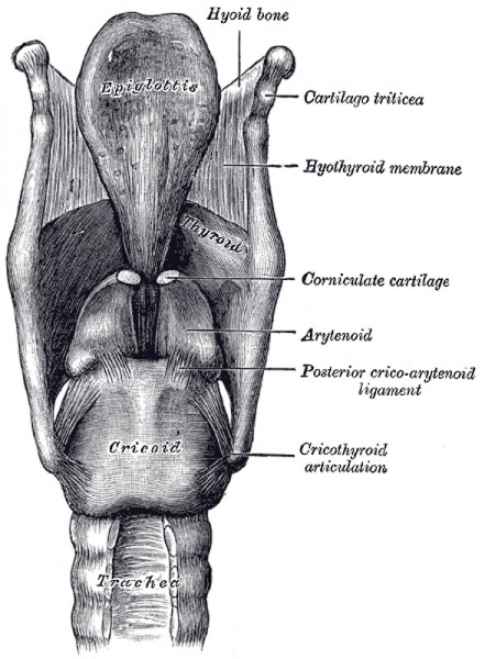
The ligaments of the larynx
Posterior view.
Ligaments
The ligaments of the larynx (Figs. 951, 952) are extrinsic, i. e., those connecting the thyroid cartilage and epiglottis with the hyoid bone, and the cricoid cartilage with the trachea; and intrinsic, those which connect the several cartilages of the larynx to each other.
Extrinsic Ligaments
The ligaments connecting the thyroid cartilage with the hyoid bone are the hyothyroid membrane, and a middle and two lateral hyothyroid ligaments.
The Hyothyroid Membrane (membrana hyothyreoidea; thyrohyoid membrane) is a broad, fibro-elastic layer, attached below to the upper border of the thyroid cartilage and to the front of its superior cornu, and above to the upper margin of the posterior surface of the body and greater cornua of the hyoid bone, thus passing behind the posterior surface of the body of the hyoid, and being separated from it by a mucous bursa, which facilitates the upward movement of the larynx during deglutition. Its middle thicker part is termed the middle hyothyroid ligament (ligamentum hyothyreoideum medium; middle thyrohyoid ligament), its lateral thinner portions are pierced by the superior laryngeal vessels and the internal branch of the superior laryngeal nerve. Its anterior surface is in relation with the Thyreohyoideus, Sternohyoideus, and Omohyoideus, and with the body of the hyoid bone.
The Lateral Hyothyroid Ligament (ligamentum hyothyreoideum laterale; lateral thyrohyoid ligament) is a round elastic cord, which forms the posterior border of the hyothyroid membrane and passes between the tip of the superior cornu of the thyroid cartilage and the extremity of the greater cornu of the hyoid bone. A small cartilaginous nodule (cartilago triticea), sometimes bony, is frequently found in it.
The Epiglottis is connected with the hyoid bone by an elastic band, the hyoepiglottic ligament (ligamentum hyoepiglotticum), which extends from the anterior surface of the epiglottis to the upper border of the body of the hyoid bone. The glossoepiglottic folds of mucous membrane (page 1075) may also be considered as extrinsic ligaments of the epiglottis.
The Cricotracheal Ligament (ligamentum cricotracheale) connects the cricoid cartilage with the first ring of the trachea. It resembles the fibrous membrane which connects the cartilaginous rings of the trachea to each other.
Intrinsic Ligaments
Beneath the mucous membrane of the larynx is a broad sheet of fibrous tissue containing many elastic fibers, and termed the elastic membrane of the larynx. It is subdivided on either side by the interval between the ventricular and vocal ligaments, the upper portion extends between the arytenoid cartilage and the epiglottis and is often poorly defined; the lower part is a well-marked membrane forming, with its fellow of the opposite side, the conus elasticus which connects the thyroid, cricoid, and arytenoid cartilages to one another. In addition the joints between the individual cartilages are provided with ligaments.
The Conus Elasticus (cricothyroid membrane) is composed mainly of yellow elastic tissue. It consists of an anterior and two lateral portions. The anterior part or middle cricothyroid ligament (ligamentum cricothyreoideum medium; central part of cricothyroid membrane) is thick and strong, narrow above and broad below. It connects together the front parts of the contiguous margins of the thyroid and cricoid cartilages. It is overlapped on either side by the Cricothyreoideus, but between these is subcutaneous; it is crossed horizontally by a small anastomotic arterial arch, formed by the junction of the two cricothyroid arteries, branches of which pierce it. The lateral portions are thinner and lie close under the mucous membrane of the larynx; they extend from the superior border of the cricoid cartilage to the inferior margin of the vocal ligaments, with which they are continuous. These ligaments may therefore be regarded as the free borders of the lateral portions of the conus elasticus, and extend from the vocal processes of the arytenoid cartilages to the angle of the thyroid cartilage about midway between its upper and lower borders.
An articular capsule, strengthened posteriorly by a well-marked fibrous band, encloses the articulation of the inferior cornu of the thyroid with the cricoid cartilage on either side.
Each arytenoid cartilage is connected to the cricoid by a capsule and a posterior cricoarytenoid ligament. The capsule (capsula articularis cricoarytenoidea) is thin and loose, and is attached to the margins of the articular surfaces. The posterior cricoarytenoid ligament (ligamentum cricoarytenoideum posterius) extends from the cricoid to the medial and back part of the base of the arytenoid.
The thyroepiglottic ligament (ligamentum thyreoepiglotticum) is a long, slender, elastic cord which connects the stem of the epiglottis with the angle of the thyroid cartilage, immediately beneath the superior thyroid notch, above the attachment of the ventricular ligaments.
(Text modified from Gray's 1918 Anatomy)
- Larynx Image Links: All cartilages of the larynx | Epiglottis cartilage | Thyroid cartilage | Cricoid cartilage | Arytenoid cartilage | Larynx ligaments anterior | Larynx ligaments posterior | Larynx sagittal section | Larynx and upper trachea | Larynx entrance | Larynx interior | Larynx muscular attachments | Larynx muscles 1 | Larynx muscles 2 | Larynx muscles 3 | Cartilage Development | Respiratory System Development
- Gray's Images: Development | Lymphatic | Neural | Vision | Hearing | Somatosensory | Integumentary | Respiratory | Gastrointestinal | Urogenital | Endocrine | Surface Anatomy | iBook | Historic Disclaimer
| Historic Disclaimer - information about historic embryology pages |
|---|
| Pages where the terms "Historic" (textbooks, papers, people, recommendations) appear on this site, and sections within pages where this disclaimer appears, indicate that the content and scientific understanding are specific to the time of publication. This means that while some scientific descriptions are still accurate, the terminology and interpretation of the developmental mechanisms reflect the understanding at the time of original publication and those of the preceding periods, these terms, interpretations and recommendations may not reflect our current scientific understanding. (More? Embryology History | Historic Embryology Papers) |
| iBook - Gray's Embryology | |
|---|---|

|
|
Reference
Gray H. Anatomy of the human body. (1918) Philadelphia: Lea & Febiger.
Cite this page: Hill, M.A. (2024, April 27) Embryology Gray0952.jpg. Retrieved from https://embryology.med.unsw.edu.au/embryology/index.php/File:Gray0952.jpg
- © Dr Mark Hill 2024, UNSW Embryology ISBN: 978 0 7334 2609 4 - UNSW CRICOS Provider Code No. 00098G
File history
Click on a date/time to view the file as it appeared at that time.
| Date/Time | Thumbnail | Dimensions | User | Comment | |
|---|---|---|---|---|---|
| current | 09:56, 21 August 2012 |  | 588 × 800 (102 KB) | Z8600021 (talk | contribs) | ==The ligaments of the larynx== Posterior view. ===Ligaments=== The ligaments of the larynx (Figs. 951, 952) are extrinsic, i. e., those connecting the thyroid cartilage and epiglottis with the hyoid bone, and the cricoid cartilage with the trachea; an |
You cannot overwrite this file.
File usage
The following 4 pages use this file:
