File:Gray0950 thyroid cartilage.jpg
Gray0950_thyroid_cartilage.jpg (600 × 500 pixels, file size: 54 KB, MIME type: image/jpeg)
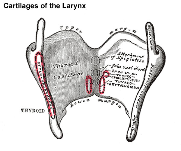
Thyroid Cartilage
(cartilago thyreoidea) is the largest cartilage of the larynx. It consists of two laminæ the anterior borders of which are fused with each other at an acute angle in the middle line of the neck, and form a subcutaneous projection named the laryngeal prominence (pomum Adami). This prominence is most distinct at its upper part, and is larger in the male than in the female. Immediately above it the laminæ are separated by a V-shaped notch, the superior thyroid notch. The laminæ are irregularly quadrilateral in shape, and their posterior angles are prolonged into processes termed the superior and inferior cornua.
The outer surface of each lamina presents an oblique line which runs downward and forward from the superior thyroid tubercle situated near the root of the superior cornu, to the inferior thyroid tubercle on the lower border. This line gives attachment to the Sternothyreoideus, Thyreohyoideus, and Constrictor pharyngis inferior.
The inner surface is smooth; above and behind, it is slightly concave and covered by mucous membrane. In front, in the angle formed by the junction of the laminæ, are attached the stem of the epiglottis, the ventricular and vocal ligaments, the Thyreoarytænoidei, Thyreoepiglottici and Vocales muscles, and the thyroepiglottic ligament.
The upper border is concave behind and convex in front; it gives attachment to the corresponding half of the hyothyroid membrane.
The lower border is concave behind, and nearly straight in front, the two parts being separated by the inferior thyroid tubercle. A small part of it in and near the middle line is connected to the cricoid cartilage by the middle cricothyroid ligament.
The posterior border, thick and rounded, receives the insertions of the Stylopharyngeus and Pharyngopalatinus. It ends above, in the superior cornu, and below, in the inferior cornu. The superior cornu is long and narrow, directed upward, backward, and medialward, and ends in a conical extremity, which gives attachment to the lateral hyothyroid ligament. The inferior cornu is short and thick; it is directed downward, with a slight inclination forward and medialward, and presents, on the medial side of its tip, a small oval articular facet for articulation with the side of the cricoid cartilage.
During infancy the laminæ of the thyroid cartilage are joined to each other by a narrow, lozenge-shaped strip, named the intrathyroid cartilage. This strip extends from the upper to the lower border of the cartilage in the middle line, and is distinguished from the laminæ by being more transparent and more flexible.
(Text modified from Gray's 1918 Anatomy)
- Larynx Image Links: All cartilages of the larynx | Epiglottis cartilage | Thyroid cartilage | Cricoid cartilage | Arytenoid cartilage | Larynx ligaments anterior | Larynx ligaments posterior | Larynx sagittal section | Larynx and upper trachea | Larynx entrance | Larynx interior | Larynx muscular attachments | Larynx muscles 1 | Larynx muscles 2 | Larynx muscles 3 | Cartilage Development | Respiratory System Development
- Gray's Images: Development | Lymphatic | Neural | Vision | Hearing | Somatosensory | Integumentary | Respiratory | Gastrointestinal | Urogenital | Endocrine | Surface Anatomy | iBook | Historic Disclaimer
| Historic Disclaimer - information about historic embryology pages |
|---|
 |
| iBook - Gray's Embryology | |
|---|---|

|
|
Reference
Gray H. Anatomy of the human body. (1918) Philadelphia: Lea & Febiger.
Cite this page: Hill, M.A. (2024, April 27) Embryology Gray0950 thyroid cartilage.jpg. Retrieved from https://embryology.med.unsw.edu.au/embryology/index.php/File:Gray0950_thyroid_cartilage.jpg
- © Dr Mark Hill 2024, UNSW Embryology ISBN: 978 0 7334 2609 4 - UNSW CRICOS Provider Code No. 00098G
File history
Click on a date/time to view the file as it appeared at that time.
| Date/Time | Thumbnail | Dimensions | User | Comment | |
|---|---|---|---|---|---|
| current | 09:28, 21 August 2012 | | 600 × 500 (54 KB) | Z8600021 (talk | contribs) | ==The Cartilages of the Larynx== Posterior view. The Cartilages of the Larynx (cartilagines laryngis) (Fig. 950) are nine in number, three single and three paired, as follows: * Thyroid. * Two Corniculate. * Cricoid. * Two Cuneiform. * Two Arytenoid. |
You cannot overwrite this file.
File usage
The following 4 pages use this file: