File:Gray0950 epiglottis cartilage.jpg
Gray0950_epiglottis_cartilage.jpg (600 × 500 pixels, file size: 24 KB, MIME type: image/jpeg)
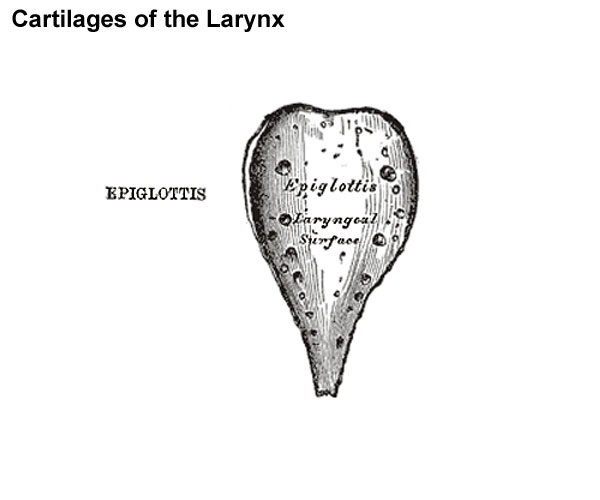
Epiglottis
Posterior view.
(cartilago epiglottica) is a thin lamella of fibrocartilage of a yellowish color, shaped like a leaf, and projecting obliquely upward behind the root of the tongue, in front of the entrance to the larynx. The free extremity is broad and rounded; the attached part or stem is long, narrow, and connected by the thyroepiglottic ligament to the angle formed by the two laminæ of the thyroid cartilage, a short distance below the superior thyroid notch. The lower part of its anterior surface is connected to the upper border of the body of the hyoid bone by an elastic ligamentous band, the hyoepiglottic ligament.
The anterior or lingual surface is curved forward, and covered on its upper, free part by mucous membrane which is reflected on to the sides and root of the tongue, forming a median and two lateral glossoepiglottic folds; the lateral folds are partly attached to the wall of the pharynx. The depressions between the epiglottis and the root of the tongue, on either side of the median fold, are named the valleculæ. The lower part of the anterior surface lies behind the hyoid bone, the hyothyroid membrane, and upper part of the thyroid cartilage, but is separated from these structures by a mass of fatty tissue.
The posterior or laryngeal surface is smooth, concave from side to side, concavo-convex from above downward; its lower part projects backward as an elevation, the tubercle or cushion. When the mucous membrane is removed, the surface of the cartilage is seen to be indented by a number of small pits, in which mucous glands are lodged. To its sides the aryepiglottic folds are attached.
(Text modified from Gray's 1918 Anatomy)
- Larynx Image Links: All cartilages of the larynx | Epiglottis cartilage | Thyroid cartilage | Cricoid cartilage | Arytenoid cartilage | Larynx ligaments anterior | Larynx ligaments posterior | Larynx sagittal section | Larynx and upper trachea | Larynx entrance | Larynx interior | Larynx muscular attachments | Larynx muscles 1 | Larynx muscles 2 | Larynx muscles 3 | Cartilage Development | Respiratory System Development
- Gray's Images: Development | Lymphatic | Neural | Vision | Hearing | Somatosensory | Integumentary | Respiratory | Gastrointestinal | Urogenital | Endocrine | Surface Anatomy | iBook | Historic Disclaimer
| Historic Disclaimer - information about historic embryology pages |
|---|
 |
| iBook - Gray's Embryology | |
|---|---|

|
|
Reference
Gray H. Anatomy of the human body. (1918) Philadelphia: Lea & Febiger.
Cite this page: Hill, M.A. (2024, April 27) Embryology Gray0950 epiglottis cartilage.jpg. Retrieved from https://embryology.med.unsw.edu.au/embryology/index.php/File:Gray0950_epiglottis_cartilage.jpg
- © Dr Mark Hill 2024, UNSW Embryology ISBN: 978 0 7334 2609 4 - UNSW CRICOS Provider Code No. 00098G
File history
Click on a date/time to view the file as it appeared at that time.
| Date/Time | Thumbnail | Dimensions | User | Comment | |
|---|---|---|---|---|---|
| current | 09:27, 21 August 2012 | | 600 × 500 (24 KB) | Z8600021 (talk | contribs) | ==Epiglottis== Posterior view. (cartilago epiglottica) is a thin lamella of fibrocartilage of a yellowish color, shaped like a leaf, and projecting obliquely upward behind the root of the tongue, in front of the entrance to the larynx. The free extremit |
You cannot overwrite this file.
File usage
The following 4 pages use this file: