File:Gray0950 cricoid cartilage.jpg
Gray0950_cricoid_cartilage.jpg (600 × 500 pixels, file size: 42 KB, MIME type: image/jpeg)
Cricoid Cartilage
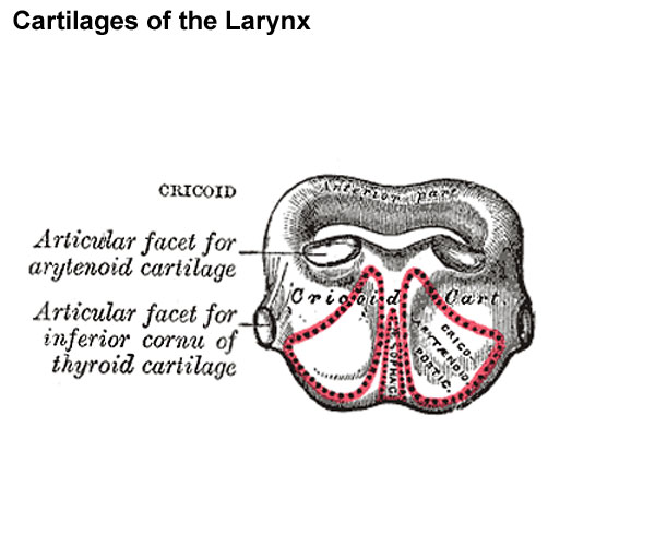
Posterior view.
(cartilago cricoidea) is smaller, but thicker and stronger than the thyroid, and forms the lower and posterior parts of the wall of the larynx. It consists of two parts: a posterior quadrate lamina, and a narrow anterior arch, one-fourth or one-fifth of the depth of the lamina.
The lamina (lamina cartilaginis cricoideæ; posterior portion) is deep and broad, and measures from above downward about 2 or 3 cm.; on its posterior surface, in the middle line, is a vertical ridge to the lower part of which are attached the longitudinal fibers of the esophagus; and on either side of this a broad depression for the Cricoarytænoideus posterior.
The arch (arcus cartilaginis cricoideæ; anterior portion) is narrow and convex, and measures vertically from 5 to 7 mm.; it affords attachment externally in front and at the sides to the Cricothyreiodei, and behind, to part of the Constrictor pharyngis inferior.
On either side, at the junction of the lamina with the arch, is a small round articular surface, for articulation with the inferior cornu of the thyroid cartilage.
The lower border of the cricoid cartilage is horizontal, and connected to the highest ring of the trachea by the cricotracheal ligament.
The upper border runs obliquely upward and backward, owing to the great depth of the lamina. It gives attachment, in front, to the middle cricothyroid ligament; at the side, to the conus elasticus and the Cricoarytænoidei laterales; behind, it presents, in the middle, a shallow notch, and on either side of this is a smooth, oval, convex surface, directed upward and lateralward, for articulation with the base of an arytenoid cartilage.
The inner surface of the cricoid cartilage is smooth, and lined by mucous membrane.
(Text modified from Gray's 1918 Anatomy)
- Larynx Image Links: All cartilages of the larynx | Epiglottis cartilage | Thyroid cartilage | Cricoid cartilage | Arytenoid cartilage | Larynx ligaments anterior | Larynx ligaments posterior | Larynx sagittal section | Larynx and upper trachea | Larynx entrance | Larynx interior | Larynx muscular attachments | Larynx muscles 1 | Larynx muscles 2 | Larynx muscles 3 | Cartilage Development | Respiratory System Development
- Gray's Images: Development | Lymphatic | Neural | Vision | Hearing | Somatosensory | Integumentary | Respiratory | Gastrointestinal | Urogenital | Endocrine | Surface Anatomy | iBook | Historic Disclaimer
| Historic Disclaimer - information about historic embryology pages |
|---|
| Pages where the terms "Historic" (textbooks, papers, people, recommendations) appear on this site, and sections within pages where this disclaimer appears, indicate that the content and scientific understanding are specific to the time of publication. This means that while some scientific descriptions are still accurate, the terminology and interpretation of the developmental mechanisms reflect the understanding at the time of original publication and those of the preceding periods, these terms, interpretations and recommendations may not reflect our current scientific understanding. (More? Embryology History | Historic Embryology Papers) |
| iBook - Gray's Embryology | |
|---|---|

|
|
Reference
Gray H. Anatomy of the human body. (1918) Philadelphia: Lea & Febiger.
Cite this page: Hill, M.A. (2024, April 27) Embryology Gray0950 cricoid cartilage.jpg. Retrieved from https://embryology.med.unsw.edu.au/embryology/index.php/File:Gray0950_cricoid_cartilage.jpg
- © Dr Mark Hill 2024, UNSW Embryology ISBN: 978 0 7334 2609 4 - UNSW CRICOS Provider Code No. 00098G
File history
Click on a date/time to view the file as it appeared at that time.
| Date/Time | Thumbnail | Dimensions | User | Comment | |
|---|---|---|---|---|---|
| current | 09:30, 21 August 2012 |  | 600 × 500 (42 KB) | Z8600021 (talk | contribs) |
You cannot overwrite this file.
File usage
The following 4 pages use this file:

