File:Gray0950 arytenoid cartilage.jpg
Gray0950_arytenoid_cartilage.jpg (600 × 500 pixels, file size: 39 KB, MIME type: image/jpeg)
Arytenoid Cartilages
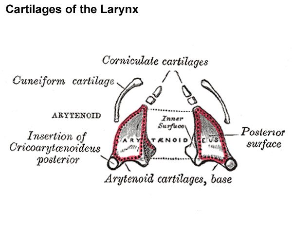
Posterior view.
(cartilagines arytænoideæ) are two in number, and situated at the upper border of the lamina of the cricoid cartilage, at the back of the larynx. Each is pyramidal in form, and has three surfaces, a base, and an apex.
The posterior surface is a triangular, smooth, concave, and gives attachment to the Arytænoidei obliquus and transversus.
The antero-lateral surface is somewhat convex and rough. On it, near the apex of the cartilage, is a rounded elevation (colliculus) from which a ridge (crista arcuata) curves at first backward and then downward and forward to the vocal process. The lower part of this crest intervenes between two depressions or foveæ, an upper, triangular, and a lower oblong in shape; the latter gives attachment to the Vocalis muscle.
The medial surface is narrow, smooth, and flattened, covered by mucous membrane, and forms the lateral boundary of the intercartilaginous part of the rima glottidis.
The base of each cartilage is broad, and on it is a concave smooth surface, for articulation with the cricoid cartilage. Its lateral angle is short, rounded, and prominent; it projects backward and lateralward, and is termed the muscular process; it gives insertion to the Cricoarytænoideus posterior behind, and to the Cricoarytænoideus lateralis in front. Its anterior angle, also prominent, but more pointed, projects horizontally forward; it gives attachment to the vocal ligament, and is called the vocal process.
The apex of each cartilage is pointed, curved backward and medialward, and surmounted by a small conical, cartilaginous nodule, the corniculate cartilage.
Corniculate Cartilages
(cartilagines corniculatæ; cartilages of Santorini) are two small conical nodules consisting of yellow elastic cartilage, which articulate with the summits of the arytenoid cartilages and serve to prolong them backward and medialward. They are situated in the posterior parts of the aryepiglottic folds of mucous membrane, and are sometimes fused with the arytenoid cartilages.
Cuneiform Cartilages
(cartilagines cuneiformes; cartilages of Wrisberg) are two small, elongated pieces of yellow elastic cartilage, placed one on either side, in the aryepiglottic fold, where they give rise to small whitish elevations on the surface of the mucous membrane, just in front of the arytenoid cartilages.
(Text modified from Gray's 1918 Anatomy)
- Larynx Image Links: All cartilages of the larynx | Epiglottis cartilage | Thyroid cartilage | Cricoid cartilage | Arytenoid cartilage | Larynx ligaments anterior | Larynx ligaments posterior | Larynx sagittal section | Larynx and upper trachea | Larynx entrance | Larynx interior | Larynx muscular attachments | Larynx muscles 1 | Larynx muscles 2 | Larynx muscles 3 | Cartilage Development | Respiratory System Development
- Gray's Images: Development | Lymphatic | Neural | Vision | Hearing | Somatosensory | Integumentary | Respiratory | Gastrointestinal | Urogenital | Endocrine | Surface Anatomy | iBook | Historic Disclaimer
| Historic Disclaimer - information about historic embryology pages |
|---|
| Pages where the terms "Historic" (textbooks, papers, people, recommendations) appear on this site, and sections within pages where this disclaimer appears, indicate that the content and scientific understanding are specific to the time of publication. This means that while some scientific descriptions are still accurate, the terminology and interpretation of the developmental mechanisms reflect the understanding at the time of original publication and those of the preceding periods, these terms, interpretations and recommendations may not reflect our current scientific understanding. (More? Embryology History | Historic Embryology Papers) |
| iBook - Gray's Embryology | |
|---|---|

|
|
Reference
Gray H. Anatomy of the human body. (1918) Philadelphia: Lea & Febiger.
Cite this page: Hill, M.A. (2024, April 27) Embryology Gray0950 arytenoid cartilage.jpg. Retrieved from https://embryology.med.unsw.edu.au/embryology/index.php/File:Gray0950_arytenoid_cartilage.jpg
- © Dr Mark Hill 2024, UNSW Embryology ISBN: 978 0 7334 2609 4 - UNSW CRICOS Provider Code No. 00098G
File history
Click on a date/time to view the file as it appeared at that time.
| Date/Time | Thumbnail | Dimensions | User | Comment | |
|---|---|---|---|---|---|
| current | 09:29, 21 August 2012 |  | 600 × 500 (39 KB) | Z8600021 (talk | contribs) | ==The Cartilages of the Larynx== Posterior view. The Cartilages of the Larynx (cartilagines laryngis) (Fig. 950) are nine in number, three single and three paired, as follows: * Thyroid. * Two Corniculate. * Cricoid. * Two Cuneiform. * Two Arytenoid. |
You cannot overwrite this file.
File usage
The following 4 pages use this file:

