File:Gray0950.jpg
Original file (500 × 1,000 pixels, file size: 106 KB, MIME type: image/jpeg)
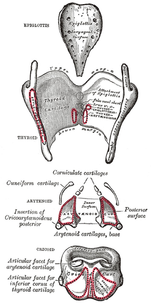
The Cartilages of the Larynx
Posterior view.
The Cartilages of the Larynx (cartilagines laryngis) (Fig. 950) are nine in number, three single and three paired, as follows:
- Thyroid
- Two Corniculate.
- Cricoid
- Two Cuneiform.
- Two Arytenoid
Thyroid Cartilage
(cartilago thyreoidea) is the largest cartilage of the larynx. It consists of two laminæ the anterior borders of which are fused with each other at an acute angle in the middle line of the neck, and form a subcutaneous projection named the laryngeal prominence (pomum Adami). This prominence is most distinct at its upper part, and is larger in the male than in the female. Immediately above it the laminæ are separated by a V-shaped notch, the superior thyroid notch. The laminæ are irregularly quadrilateral in shape, and their posterior angles are prolonged into processes termed the superior and inferior cornua.
The outer surface of each lamina presents an oblique line which runs downward and forward from the superior thyroid tubercle situated near the root of the superior cornu, to the inferior thyroid tubercle on the lower border. This line gives attachment to the Sternothyreoideus, Thyreohyoideus, and Constrictor pharyngis inferior.
The inner surface is smooth; above and behind, it is slightly concave and covered by mucous membrane. In front, in the angle formed by the junction of the laminæ, are attached the stem of the epiglottis, the ventricular and vocal ligaments, the Thyreoarytænoidei, Thyreoepiglottici and Vocales muscles, and the thyroepiglottic ligament.
The upper border is concave behind and convex in front; it gives attachment to the corresponding half of the hyothyroid membrane.
The lower border is concave behind, and nearly straight in front, the two parts being separated by the inferior thyroid tubercle. A small part of it in and near the middle line is connected to the cricoid cartilage by the middle cricothyroid ligament.
The posterior border, thick and rounded, receives the insertions of the Stylopharyngeus and Pharyngopalatinus. It ends above, in the superior cornu, and below, in the inferior cornu. The superior cornu is long and narrow, directed upward, backward, and medialward, and ends in a conical extremity, which gives attachment to the lateral hyothyroid ligament. The inferior cornu is short and thick; it is directed downward, with a slight inclination forward and medialward, and presents, on the medial side of its tip, a small oval articular facet for articulation with the side of the cricoid cartilage.
During infancy the laminæ of the thyroid cartilage are joined to each other by a narrow, lozenge-shaped strip, named the intrathyroid cartilage. This strip extends from the upper to the lower border of the cartilage in the middle line, and is distinguished from the laminæ by being more transparent and more flexible.
Cricoid Cartilage
(cartilago cricoidea) is smaller, but thicker and stronger than the thyroid, and forms the lower and posterior parts of the wall of the larynx. It consists of two parts: a posterior quadrate lamina, and a narrow anterior arch, one-fourth or one-fifth of the depth of the lamina.
The lamina (lamina cartilaginis cricoideæ; posterior portion) is deep and broad, and measures from above downward about 2 or 3 cm.; on its posterior surface, in the middle line, is a vertical ridge to the lower part of which are attached the longitudinal fibers of the esophagus; and on either side of this a broad depression for the Cricoarytænoideus posterior.
The arch (arcus cartilaginis cricoideæ; anterior portion) is narrow and convex, and measures vertically from 5 to 7 mm.; it affords attachment externally in front and at the sides to the Cricothyreiodei, and behind, to part of the Constrictor pharyngis inferior.
On either side, at the junction of the lamina with the arch, is a small round articular surface, for articulation with the inferior cornu of the thyroid cartilage.
The lower border of the cricoid cartilage is horizontal, and connected to the highest ring of the trachea by the cricotracheal ligament.
The upper border runs obliquely upward and backward, owing to the great depth of the lamina. It gives attachment, in front, to the middle cricothyroid ligament; at the side, to the conus elasticus and the Cricoarytænoidei laterales; behind, it presents, in the middle, a shallow notch, and on either side of this is a smooth, oval, convex surface, directed upward and lateralward, for articulation with the base of an arytenoid cartilage.
The inner surface of the cricoid cartilage is smooth, and lined by mucous membrane.
Arytenoid Cartilages
(cartilagines arytænoideæ) are two in number, and situated at the upper border of the lamina of the cricoid cartilage, at the back of the larynx. Each is pyramidal in form, and has three surfaces, a base, and an apex.
The posterior surface is a triangular, smooth, concave, and gives attachment to the Arytænoidei obliquus and transversus.
The antero-lateral surface is somewhat convex and rough. On it, near the apex of the cartilage, is a rounded elevation (colliculus) from which a ridge (crista arcuata) curves at first backward and then downward and forward to the vocal process. The lower part of this crest intervenes between two depressions or foveæ, an upper, triangular, and a lower oblong in shape; the latter gives attachment to the Vocalis muscle.
The medial surface is narrow, smooth, and flattened, covered by mucous membrane, and forms the lateral boundary of the intercartilaginous part of the rima glottidis.
The base of each cartilage is broad, and on it is a concave smooth surface, for articulation with the cricoid cartilage. Its lateral angle is short, rounded, and prominent; it projects backward and lateralward, and is termed the muscular process; it gives insertion to the Cricoarytænoideus posterior behind, and to the Cricoarytænoideus lateralis in front. Its anterior angle, also prominent, but more pointed, projects horizontally forward; it gives attachment to the vocal ligament, and is called the vocal process.
The apex of each cartilage is pointed, curved backward and medialward, and surmounted by a small conical, cartilaginous nodule, the corniculate cartilage.
Corniculate Cartilages
(cartilagines corniculatæ; cartilages of Santorini) are two small conical nodules consisting of yellow elastic cartilage, which articulate with the summits of the arytenoid cartilages and serve to prolong them backward and medialward. They are situated in the posterior parts of the aryepiglottic folds of mucous membrane, and are sometimes fused with the arytenoid cartilages.
Cuneiform Cartilages
(cartilagines cuneiformes; cartilages of Wrisberg) are two small, elongated pieces of yellow elastic cartilage, placed one on either side, in the aryepiglottic fold, where they give rise to small whitish elevations on the surface of the mucous membrane, just in front of the arytenoid cartilages.
Epiglottis
(cartilago epiglottica) is a thin lamella of fibrocartilage of a yellowish color, shaped like a leaf, and projecting obliquely upward behind the root of the tongue, in front of the entrance to the larynx. The free extremity is broad and rounded; the attached part or stem is long, narrow, and connected by the thyroepiglottic ligament to the angle formed by the two laminæ of the thyroid cartilage, a short distance below the superior thyroid notch. The lower part of its anterior surface is connected to the upper border of the body of the hyoid bone by an elastic ligamentous band, the hyoepiglottic ligament.
The anterior or lingual surface is curved forward, and covered on its upper, free part by mucous membrane which is reflected on to the sides and root of the tongue, forming a median and two lateral glossoepiglottic folds; the lateral folds are partly attached to the wall of the pharynx. The depressions between the epiglottis and the root of the tongue, on either side of the median fold, are named the valleculæ. The lower part of the anterior surface lies behind the hyoid bone, the hyothyroid membrane, and upper part of the thyroid cartilage, but is separated from these structures by a mass of fatty tissue.
The posterior or laryngeal surface is smooth, concave from side to side, concavo-convex from above downward; its lower part projects backward as an elevation, the tubercle or cushion. When the mucous membrane is removed, the surface of the cartilage is seen to be indented by a number of small pits, in which mucous glands are lodged. To its sides the aryepiglottic folds are attached.
(Text modified from Gray's 1918 Anatomy)
- Larynx Image Links: All cartilages of the larynx | Epiglottis cartilage | Thyroid cartilage | Cricoid cartilage | Arytenoid cartilage | Larynx ligaments anterior | Larynx ligaments posterior | Larynx sagittal section | Larynx and upper trachea | Larynx entrance | Larynx interior | Larynx muscular attachments | Larynx muscles 1 | Larynx muscles 2 | Larynx muscles 3 | Cartilage Development | Respiratory System Development
- Gray's Images: Development | Lymphatic | Neural | Vision | Hearing | Somatosensory | Integumentary | Respiratory | Gastrointestinal | Urogenital | Endocrine | Surface Anatomy | iBook | Historic Disclaimer
| Historic Disclaimer - information about historic embryology pages |
|---|
 |
| iBook - Gray's Embryology | |
|---|---|

|
|
Reference
Gray H. Anatomy of the human body. (1918) Philadelphia: Lea & Febiger.
Cite this page: Hill, M.A. (2024, April 27) Embryology Gray0950.jpg. Retrieved from https://embryology.med.unsw.edu.au/embryology/index.php/File:Gray0950.jpg
- © Dr Mark Hill 2024, UNSW Embryology ISBN: 978 0 7334 2609 4 - UNSW CRICOS Provider Code No. 00098G
File history
Click on a date/time to view the file as it appeared at that time.
| Date/Time | Thumbnail | Dimensions | User | Comment | |
|---|---|---|---|---|---|
| current | 09:05, 21 August 2012 | | 500 × 1,000 (106 KB) | Z8600021 (talk | contribs) | ==The cartilages of the larynx== Posterior view. The Cartilages of the Larynx (cartilagines laryngis) (Fig. 950) are nine in number, three single and three paired, as follows: * Thyroid. * Two Corniculate. * Cricoid. * Two Cuneiform. * Two Arytenoid. |
You cannot overwrite this file.
File usage
The following 4 pages use this file: