File:Gray0933.jpg
Gray0933.jpg (500 × 360 pixels, file size: 54 KB, MIME type: image/jpeg)
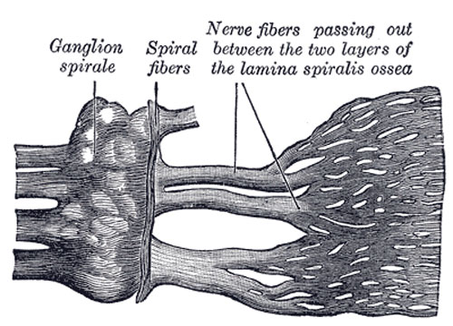
Cochlear Division of the Acoustic Nerve
Part of the cochlear division of the acoustic nerve, highly magnified. (Henle.)
The cochlear nerve (n. cochlearis) divides into numerous filaments at the base of the modiolus; those for the basal and middle coils pass through the foramina in the tractus spiralis foraminosis, those for the apical coil through the canalis centralis, and the nerves bend outward to pass between the lamellæ of the osseous spiral lamina. Occupying the spiral canal of the modiolus is the spiral ganglion of the cochlea (ganglion of Corti) (Fig. 933), consisting of bipolar nerve cells, which constitute the cells of origin of this nerve. Reaching the outer edge of the osseous spiral lamina, the fibers of the nerve pass through the foramina in the tympanic lip; some end by arborizing around the bases of the inner hair cells, while others pass between Corti’s rods and across the tunnel, to end in a similar manner in relation to the outer hair cells. The cochlear nerve gives off a vestibular branch to supply the vestibular end of the ductus cochlearis; the filaments of this branch pass through the foramina in the fossa cochlearis (page 1048).
(Text modified from Gray's 1918 Anatomy)
- Gray's Images: Development | Lymphatic | Neural | Vision | Hearing | Somatosensory | Integumentary | Respiratory | Gastrointestinal | Urogenital | Endocrine | Surface Anatomy | iBook | Historic Disclaimer
| Historic Disclaimer - information about historic embryology pages |
|---|
| Pages where the terms "Historic" (textbooks, papers, people, recommendations) appear on this site, and sections within pages where this disclaimer appears, indicate that the content and scientific understanding are specific to the time of publication. This means that while some scientific descriptions are still accurate, the terminology and interpretation of the developmental mechanisms reflect the understanding at the time of original publication and those of the preceding periods, these terms, interpretations and recommendations may not reflect our current scientific understanding. (More? Embryology History | Historic Embryology Papers) |
| iBook - Gray's Embryology | |
|---|---|

|
|
Reference
Gray H. Anatomy of the human body. (1918) Philadelphia: Lea & Febiger.
Cite this page: Hill, M.A. (2024, April 27) Embryology Gray0933.jpg. Retrieved from https://embryology.med.unsw.edu.au/embryology/index.php/File:Gray0933.jpg
- © Dr Mark Hill 2024, UNSW Embryology ISBN: 978 0 7334 2609 4 - UNSW CRICOS Provider Code No. 00098G
File history
Click on a date/time to view the file as it appeared at that time.
| Date/Time | Thumbnail | Dimensions | User | Comment | |
|---|---|---|---|---|---|
| current | 08:14, 19 August 2012 |  | 500 × 360 (54 KB) | Z8600021 (talk | contribs) | ==Cochlear Division of the Acoustic Nerve== Part of the cochlear division of the acoustic nerve, highly magnified. (Henle.) The cochlear nerve (n. cochlearis) divides into numerous filaments at the base of the modiolus; those for the basal and middle co |
You cannot overwrite this file.
File usage
The following page uses this file:

