File:Gray0929.jpg
Gray0929.jpg (700 × 287 pixels, file size: 45 KB, MIME type: image/jpeg)
Floor of Ductus Cochlearis
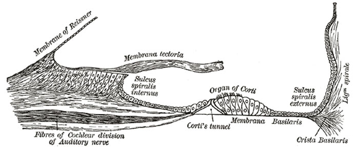
The osseous spiral lamina consists of two plates of bone, and between these are the canals for the transmission of the filaments of the acoustic nerve. On the upper plate of that part of the lamina which is outside the vestibular membrane, the periosteum is thickened to form the limbus laminæ spiralis (Fig. 929), this ends externally in a concavity, the sulcus spiralis internus, which represents, on section, the form of the letter C; the upper part, formed by the overhanging extremity of the limbus, is named the vestibular lip; the lower part, prolonged and tapering, is called the tympanic lip, and is perforated by numerous foramina for the passage of the cochlear nerves. The upper surface of the vestibular lip is intersected at right angles by a number of furrows, between which are numerous elevations; these present the appearance of teeth along the free surface and margin of the lip, and have been named by Huschke the auditory teeth (Fig. 930). The limbus is covered by a layer of what appears to be squamous epithelium, but the deeper parts of the cells with their contained nuclei occupy the intervals between the elevations and between the auditory teeth. This layer of epithelium is continuous on the one hand with that lining the sulcus spiralis internus, and on the other with that covering the under surface of the vestibular membrane.
Basilar Membrane
The basilar membrane stretches from the tympanic lip of the osseous spiral lamina to the basilar crest and consists of two parts, an inner and an outer. The inner is thin, and is named the zona arcuata: it supports the spiral organ of Corti. The outer is thicker and striated, and is termed the zona pectinata. The under surface of the membrane is covered by a layer of vascular connective tissue; one of the vessels in this tissue is somewhat larger than the rest, and is named the vas spirale; it lies below Corti’s tunnel.
(Text modified from Gray's 1918 Anatomy)
- Gray's Images: Development | Lymphatic | Neural | Vision | Hearing | Somatosensory | Integumentary | Respiratory | Gastrointestinal | Urogenital | Endocrine | Surface Anatomy | iBook | Historic Disclaimer
| Historic Disclaimer - information about historic embryology pages |
|---|
| Pages where the terms "Historic" (textbooks, papers, people, recommendations) appear on this site, and sections within pages where this disclaimer appears, indicate that the content and scientific understanding are specific to the time of publication. This means that while some scientific descriptions are still accurate, the terminology and interpretation of the developmental mechanisms reflect the understanding at the time of original publication and those of the preceding periods, these terms, interpretations and recommendations may not reflect our current scientific understanding. (More? Embryology History | Historic Embryology Papers) |
| iBook - Gray's Embryology | |
|---|---|

|
|
Reference
Gray H. Anatomy of the human body. (1918) Philadelphia: Lea & Febiger.
Cite this page: Hill, M.A. (2024, April 27) Embryology Gray0929.jpg. Retrieved from https://embryology.med.unsw.edu.au/embryology/index.php/File:Gray0929.jpg
- © Dr Mark Hill 2024, UNSW Embryology ISBN: 978 0 7334 2609 4 - UNSW CRICOS Provider Code No. 00098G
File history
Click on a date/time to view the file as it appeared at that time.
| Date/Time | Thumbnail | Dimensions | User | Comment | |
|---|---|---|---|---|---|
| current | 08:00, 19 August 2012 | 700 × 287 (45 KB) | Z8600021 (talk | contribs) | (Text modified from Gray's 1918 Anatomy) :'''Links:''' Inner Ear Development | Hearing and Balance Development | [[Anatomy_of_the_Human_Body_by_Henry_Gray|Gray's 1918 Anat |
You cannot overwrite this file.
File usage
The following page uses this file:

