File:Gray0915.jpg
Gray0915.jpg (720 × 600 pixels, file size: 94 KB, MIME type: image/jpeg)
Auditory Tube
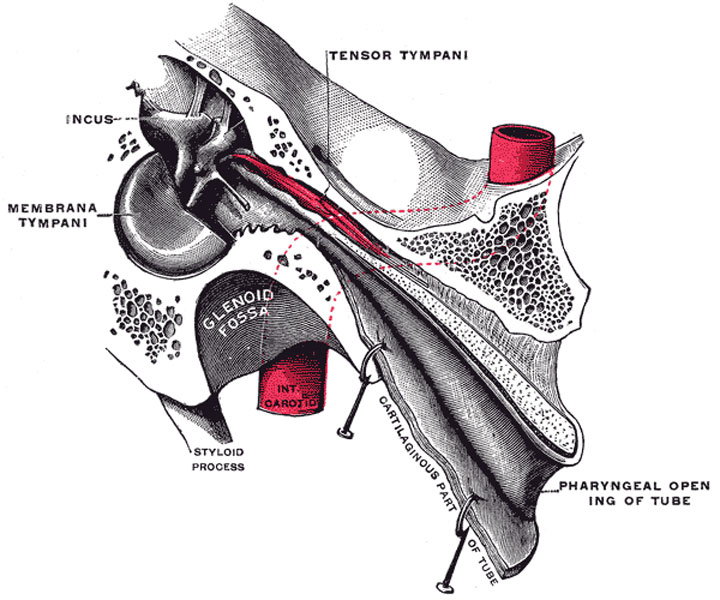
Auditory tube, laid open by a cut in its long axis. (Testut.)
Tympanic Cavity Anterior Wall
The Carotid or Anterior Wall (paries carotica) is wider above than below; it corresponds with the carotid canal, from which it is separated by a thin plate of bone perforated by the tympanic branch of the internal carotid artery, and by the deep petrosal nerve which connects the sympathetic plexus on the internal carotid artery with the tympanic plexus on the promontory. At the upper part of the anterior wall are the orifice of the semicanal for the Tensor tympani muscle and the tympanic orifice of the auditory tube, separated from each other by a thin horizontal plate of bone, the septum canalis musculotubarii. These canals run from the tympanic cavity forward and downward to the retiring angle between the squama and the petrous portion of the temporal bone.
The semicanal for the Tensor tympani (semicanalis m. tensoris tympani) is the superior and the smaller of the two; it is cylindrical and lies beneath the tegmen tympani. It extends on to the labyrinthic wall of the tympanic cavity and ends immediately above the fenestra vestibuli.
The septum canalis musculotubarii (processus cochleariformis) passes backward below this semicanal, forming its lateral wall and floor; it expands above the anterior end of the fenestra vestibuli and terminates there by curving laterally so as to form a pulley over which the tendon of the muscle passes.
The auditory tube (tuba auditiva; Eustachian tube) is the channel through which the tympanic cavity communicates with the nasal part of the pharynx. Its length is about 36 mm., and its direction is downward, forward, and medialward, forming an angle of about 45 degrees with the sagittal plane and one of from 30 to 40 degrees with the horizontal plane. It is formed partly of bone, partly of cartilage and fibrous tissue (Figs. 819, 915).
The osseous portion (pars osseo tubæ auditivæ) is about 12 mm. in length. It begins in the carotid wall of the tympanic cavity, below the septum canalis musculotubarii, and, gradually narrowing, ends at the angle of junction of the squama and the petrous portion of the temporal bone, its extremity presenting a jagged margin which serves for the attachment of the cartilaginous portion.
The cartilaginous portion (pars cartilaginea tubæ auditivæ), about 24 mm. in length, is formed of a triangular plate of elastic fibrocartilage, the apex of which is attached to the margin of the medial end of the osseous portion of the tube, while its base lies directly under the mucous membrane of the nasal part of the pharynx, where it forms an elevation, the torus tubarius or cushion, behind the pharyngeal orifice of the tube. The upper edge of the cartilage is curled upon itself, being bent laterally so as to present on transverse section the appearance of a hook; a groove or furrow is thus produced, which is open below and laterally, and this part of the canal is completed by fibrous membrane. The cartilage lies in a groove between the petrous part of the temporal and the great wing of the sphenoid; this groove ends opposite the middle of the medial pterygoid plate. The cartilaginous and bony portions of the tube are not in the same plane, the former inclining downward a little more than the latter. The diameter of the tube is not uniform throughout, being greatest at the pharyngeal orifice, least at the junction of the bony and cartilaginous portions, and again increased toward the tympanic cavity; the narrowest part of the tube is termed the isthmus. The position and relations of the pharyngeal orifice are described with the nasal part of the pharynx. The mucous membrane of the tube is continuous in front with that of the nasal part of the pharynx, and behind with that of the tympanic cavity; it is covered with ciliated epithelium and is thin in the osseous portion, while in the cartilaginous portion it contains many mucous glands and near the pharyngeal orifice a considerable amount of adenoid tissue, which has been named by Gerlach the tube tonsil. The tube is opened during deglutition by the Salpingopharyngeus and Dilatator tubæ. The latter arises from the hook of the cartilage and from the membranous part of the tube, and blends below with the Tensor veli palatini.
(Text modified from Gray's 1918 Anatomy)
- Gray's Images: Development | Lymphatic | Neural | Vision | Hearing | Somatosensory | Integumentary | Respiratory | Gastrointestinal | Urogenital | Endocrine | Surface Anatomy | iBook | Historic Disclaimer
| Historic Disclaimer - information about historic embryology pages |
|---|
| Pages where the terms "Historic" (textbooks, papers, people, recommendations) appear on this site, and sections within pages where this disclaimer appears, indicate that the content and scientific understanding are specific to the time of publication. This means that while some scientific descriptions are still accurate, the terminology and interpretation of the developmental mechanisms reflect the understanding at the time of original publication and those of the preceding periods, these terms, interpretations and recommendations may not reflect our current scientific understanding. (More? Embryology History | Historic Embryology Papers) |
| iBook - Gray's Embryology | |
|---|---|

|
|
Reference
Gray H. Anatomy of the human body. (1918) Philadelphia: Lea & Febiger.
Cite this page: Hill, M.A. (2024, April 27) Embryology Gray0915.jpg. Retrieved from https://embryology.med.unsw.edu.au/embryology/index.php/File:Gray0915.jpg
- © Dr Mark Hill 2024, UNSW Embryology ISBN: 978 0 7334 2609 4 - UNSW CRICOS Provider Code No. 00098G
File history
Click on a date/time to view the file as it appeared at that time.
| Date/Time | Thumbnail | Dimensions | User | Comment | |
|---|---|---|---|---|---|
| current | 07:13, 19 August 2012 |  | 720 × 600 (94 KB) | Z8600021 (talk | contribs) |
You cannot overwrite this file.
File usage
The following page uses this file:

