File:Gray0914.jpg
Gray0914.jpg (708 × 500 pixels, file size: 102 KB, MIME type: image/jpeg)
Right Tympanic Cavity Walls
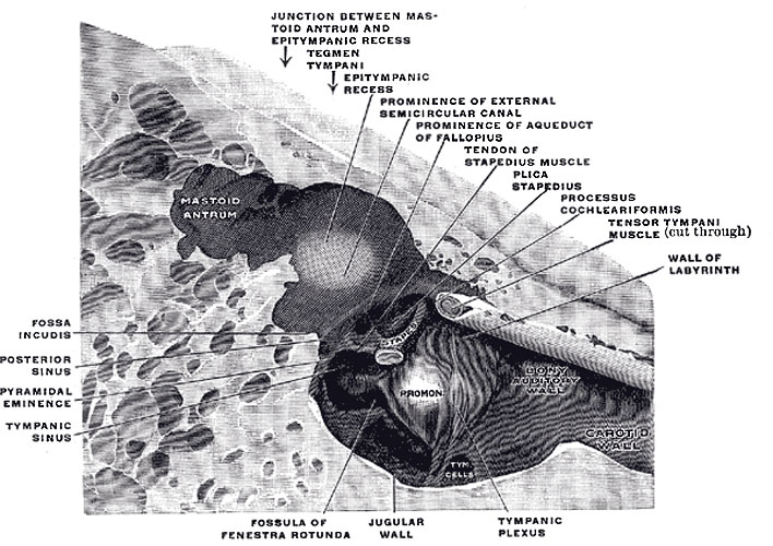
The medial wall and part of the posterior and anterior walls of the right tympanic cavity, lateral view. (Spalteholz.)
Medial Wall
The Labyrinthic or Medial Wall (paries labyrinthica; inner wall) (Fig. 913) is vertical in direction, and presents for examination the fenestræ vestibuli and cochleæ, the promontory, and the prominence of the facial canal.
The fenestra vestibuli (fenestra ovalis) is a reniform opening leading from the tympanic cavity into the vestibule of the internal ear; its long diameter is horizontal, and its convex border is upward. In the recent state it is occupied by the base of the stapes, the circumference of which is fixed by the annular ligament to the margin of the foramen.
The fenestra cochleæ (fenestra rotunda) is situated below and a little behind the fenestra vestibuli, from which it is separated by a rounded elevation, the promontory. It is placed at the bottom of a funnel-shaped depression and, in the macerated bone, leads into the cochlea of the internal ear; in the fresh state it is closed by a membrane, the secondary tympanic membrane, which is concave toward the tympanic cavity, convex toward the cochlea. This membrane consists of three layers: an external, or mucous, derived from the mucous lining of the tympanic cavity; an internal, from the lining membrane of the cochlea; and an intermediate, or fibrous layer.
The promontory (promontorium) is a rounded hollow prominence, formed by the projection outward of the first turn of the cochlea; it is placed between the fenestræ, and is furrowed on its surface by small grooves, for the lodgement of branches of the tympanic plexus. A minute spicule of bone frequently connects the promontory to the pyramidal eminence.
The prominence of the facial canal (prominentia canalis facialis; prominence of aqueduct of Fallopius) indicates the position of the bony canal in which the facial nerve is contained; this canal traverses the labyrinthic wall of the tympanic cavity above the fenestra vestibuli, and behind that opening curves nearly vertically downward along the mastoid wall.
Posterior Wall
The mastoid or posterior wall (paries mastoidea) is wider above than below, and presents for examination the entrance to the tympanic antrum, the pyramidal eminence, and the fossa incudis.
The entrance to the antrum is a large irregular aperture, which leads backward from the epitympanic recess into a considerable air space, named the tympanic or mastoid antrum (see page 142). The antrum communicates behind and below with the mastoid air cells, which vary considerably in number, size, and form; the antrum and mastoid air cells are lined by mucous membrane, continuous with that lining the tympanic cavity. On the medial wall of the entrance to the antrum is a rounded eminence, situated above and behind the prominence of the facial canal; it corresponds with the position of the ampullated ends of the superior and lateral semicircular canals.
The pyramidal eminence (eminentia pyramidalis; pyramid) is situated immediately behind the fenestra vestibuli, and in front of the vertical portion of the facial canal; it is hollow, and contains the Stapedius muscle; its summit projects forward toward the fenestra vestibuli, and is pierced by a small aperture which transmits the tendon of the muscle. The cavity in the pyramidal eminence is prolonged downward and backward in front of the facial canal, and communicates with it by a minute aperture which transmits a twig from the facial nerve to the Stapedius muscle.
The fossa incudis is a small depression in the lower and back part of the epitympanic recess; it lodges the short crus of the incus.
Anterior Wall
The Carotid or Anterior Wall (paries carotica) is wider above than below; it corresponds with the carotid canal, from which it is separated by a thin plate of bone perforated by the tympanic branch of the internal carotid artery, and by the deep petrosal nerve which connects the sympathetic plexus on the internal carotid artery with the tympanic plexus on the promontory. At the upper part of the anterior wall are the orifice of the semicanal for the Tensor tympani muscle and the tympanic orifice of the auditory tube, separated from each other by a thin horizontal plate of bone, the septum canalis musculotubarii. These canals run from the tympanic cavity forward and downward to the retiring angle between the squama and the petrous portion of the temporal bone.
(Text modified from Gray's 1918 Anatomy)
- Gray's Images: Development | Lymphatic | Neural | Vision | Hearing | Somatosensory | Integumentary | Respiratory | Gastrointestinal | Urogenital | Endocrine | Surface Anatomy | iBook | Historic Disclaimer
| Historic Disclaimer - information about historic embryology pages |
|---|
| Pages where the terms "Historic" (textbooks, papers, people, recommendations) appear on this site, and sections within pages where this disclaimer appears, indicate that the content and scientific understanding are specific to the time of publication. This means that while some scientific descriptions are still accurate, the terminology and interpretation of the developmental mechanisms reflect the understanding at the time of original publication and those of the preceding periods, these terms, interpretations and recommendations may not reflect our current scientific understanding. (More? Embryology History | Historic Embryology Papers) |
| iBook - Gray's Embryology | |
|---|---|

|
|
Reference
Gray H. Anatomy of the human body. (1918) Philadelphia: Lea & Febiger.
Cite this page: Hill, M.A. (2024, April 27) Embryology Gray0914.jpg. Retrieved from https://embryology.med.unsw.edu.au/embryology/index.php/File:Gray0914.jpg
- © Dr Mark Hill 2024, UNSW Embryology ISBN: 978 0 7334 2609 4 - UNSW CRICOS Provider Code No. 00098G
File history
Click on a date/time to view the file as it appeared at that time.
| Date/Time | Thumbnail | Dimensions | User | Comment | |
|---|---|---|---|---|---|
| current | 07:07, 19 August 2012 |  | 708 × 500 (102 KB) | Z8600021 (talk | contribs) | ==Right Tympanic Cavity Walls== The medial wall and part of the posterior and anterior walls of the right tympanic cavity, lateral view. (Spalteholz.) The Labyrinthic or Medial Wall (paries labyrinthica; inner wall) (Fig. 913) is vertical in direction, |
You cannot overwrite this file.
File usage
The following page uses this file:

