File:Gray0913.jpg
Gray0913.jpg (671 × 600 pixels, file size: 98 KB, MIME type: image/jpeg)
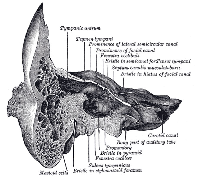
Coronal section of right temporal bone
The Labyrinthic or Medial Wall (paries labyrinthica; inner wall) (Fig. 913) is vertical in direction, and presents for examination the fenestræ vestibuli and cochleæ, the promontory, and the prominence of the facial canal.
The fenestra vestibuli (fenestra ovalis) is a reniform opening leading from the tympanic cavity into the vestibule of the internal ear; its long diameter is horizontal, and its convex border is upward. In the recent state it is occupied by the base of the stapes, the circumference of which is fixed by the annular ligament to the margin of the foramen.
The fenestra cochleæ (fenestra rotunda) is situated below and a little behind the fenestra vestibuli, from which it is separated by a rounded elevation, the promontory. It is placed at the bottom of a funnel-shaped depression and, in the macerated bone, leads into the cochlea of the internal ear; in the fresh state it is closed by a membrane, the secondary tympanic membrane, which is concave toward the tympanic cavity, convex toward the cochlea. This membrane consists of three layers: an external, or mucous, derived from the mucous lining of the tympanic cavity; an internal, from the lining membrane of the cochlea; and an intermediate, or fibrous layer.
(Text modified from Gray's 1918 Anatomy)
- Gray's Images: Development | Lymphatic | Neural | Vision | Hearing | Somatosensory | Integumentary | Respiratory | Gastrointestinal | Urogenital | Endocrine | Surface Anatomy | iBook | Historic Disclaimer
| Historic Disclaimer - information about historic embryology pages |
|---|
| Pages where the terms "Historic" (textbooks, papers, people, recommendations) appear on this site, and sections within pages where this disclaimer appears, indicate that the content and scientific understanding are specific to the time of publication. This means that while some scientific descriptions are still accurate, the terminology and interpretation of the developmental mechanisms reflect the understanding at the time of original publication and those of the preceding periods, these terms, interpretations and recommendations may not reflect our current scientific understanding. (More? Embryology History | Historic Embryology Papers) |
| iBook - Gray's Embryology | |
|---|---|

|
|
Reference
Gray H. Anatomy of the human body. (1918) Philadelphia: Lea & Febiger.
Cite this page: Hill, M.A. (2024, April 27) Embryology Gray0913.jpg. Retrieved from https://embryology.med.unsw.edu.au/embryology/index.php/File:Gray0913.jpg
- © Dr Mark Hill 2024, UNSW Embryology ISBN: 978 0 7334 2609 4 - UNSW CRICOS Provider Code No. 00098G
File history
Click on a date/time to view the file as it appeared at that time.
| Date/Time | Thumbnail | Dimensions | User | Comment | |
|---|---|---|---|---|---|
| current | 07:01, 19 August 2012 |  | 671 × 600 (98 KB) | Z8600021 (talk | contribs) | ==Coronal section of right temporal bone== (Text modified from Gray's 1918 Anatomy) :'''Links:''' Middle Ear Development | Hearing and Balance Development | [[Anatomy_of |
You cannot overwrite this file.
File usage
The following page uses this file:

