File:Gray0911.jpg
Gray0911.jpg (651 × 400 pixels, file size: 73 KB, MIME type: image/jpeg)
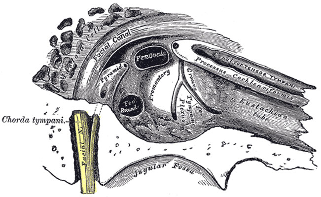
View of the inner wall of the Tympanum
The Tympanic Membrane
(membrana tympani) (Fig. 909, Fig. 910) separates the tympanic cavity from the bottom of the external acoustic meatus. It is a thin, semitransparent membrane, nearly oval in form, somewhat broader above than below, and directed very obliquely downward and inward so as to form an angle of about fifty-five degrees with the floor of the meatus. Its longest diameter is downward and forward, and measures from 9 to 10 mm.; its shortest diameter measures from 8 to 9 mm.
The greater part of its circumference is thickened, and forms a fibrocartilaginous ring which is fixed in the tympanic sulcus at the inner end of the meatus. This sulcus is deficient superiorly at the notch of Rivinus, and from the ends of this notch two bands, the anterior and posterior malleolar folds, are prolonged to the lateral process of the malleus.
The small, somewhat triangular part of the membrane situated above these folds is lax and thin, and is named the pars flaccida; in it a small orifice is sometimes seen.
The manubrium of the malleus is firmly attached to the medial surface of the membrane as far as its center, which it draws toward the tympanic cavity; the lateral surface of the membrane is thus concave, and the most depressed part of this concavity is named the umbo.
(Text modified from Gray's 1918 Anatomy)
- Gray's Images: Development | Lymphatic | Neural | Vision | Hearing | Somatosensory | Integumentary | Respiratory | Gastrointestinal | Urogenital | Endocrine | Surface Anatomy | iBook | Historic Disclaimer
| Historic Disclaimer - information about historic embryology pages |
|---|
 |
| iBook - Gray's Embryology | |
|---|---|

|
|
Reference
Gray H. Anatomy of the human body. (1918) Philadelphia: Lea & Febiger.
Cite this page: Hill, M.A. (2024, April 27) Embryology Gray0911.jpg. Retrieved from https://embryology.med.unsw.edu.au/embryology/index.php/File:Gray0911.jpg
- © Dr Mark Hill 2024, UNSW Embryology ISBN: 978 0 7334 2609 4 - UNSW CRICOS Provider Code No. 00098G
File history
Click on a date/time to view the file as it appeared at that time.
| Date/Time | Thumbnail | Dimensions | User | Comment | |
|---|---|---|---|---|---|
| current | 06:52, 19 August 2012 | | 651 × 400 (73 KB) | Z8600021 (talk | contribs) | == View of the inner wall of the Tympanum== ===The Tympanic Membrane=== (membrana tympani) (Fig. 909, Fig. 910) separates the tympanic cavity from the bottom of the external acoustic meatus. It is a thin, se |
You cannot overwrite this file.
File usage
The following page uses this file: