File:Gray0909.jpg
From Embryology
No higher resolution available.
Gray0909.jpg (600 × 437 pixels, file size: 56 KB, MIME type: image/jpeg)
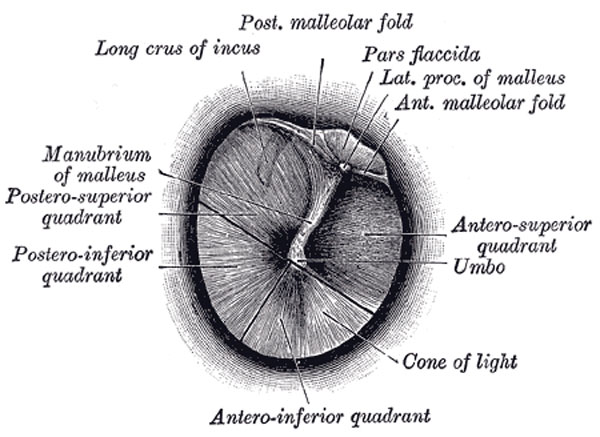
Right Tympanic Membrane
Right tympanic membrane as seen through a speculum.
The Membranous or Lateral Wall (paries membranacea; outer wall) is formed mainly by the tympanic membrane, partly by the ring of bone into which this membrane is inserted. This ring of bone is incomplete at its upper part, forming a notch (notch of Rivinus), close to which are three small apertures: the iter chordæ posterius, the petrotympanic fissure, and the iter chordæ anterius.	   5
 The iter chordæ posterius (apertura tympanica canaliculi chordæ) is situated in the angle of junction between the mastoid and membranous wall of the tympanic cavity immediately behind the tympanic membrane and on a level with the upper end of the manubrium of the malleus; it leads into a minute canal, which descends in front of the canal for the facial nerve, and ends in that canal near the stylo-mastoid foramen. Through it the chorda tympani nerve enters the tympanic cavity.	   6
 The petrotympanic fissure (fissura petrotympanica; Glaserian fissure) opens just above and in front of the ring of bone into which the tympanic membrane is inserted; in this situation it is a mere slit about 2 mm. in length. It lodges the anterior process and anterior ligament of the malleus, and gives passage to the anterior tympanic branch of the internal maxillary artery.	   7
 The iter chordæ anterius (canal of Huguier) is placed at the medial end of the petrotympanic fissure; through it the chorda tympani nerve leaves the tympanic cavity.	   8
 The Tympanic Membrane (membrana tympani) (Figs. 909, 910) separates the tympanic cavity from the bottom of the external acoustic meatus. It is a thin, semitransparent membrane, nearly oval in form, somewhat broader above than below, and directed very obliquely downward and inward so as to form an angle of about fifty-five degrees with the floor of the meatus. Its longest diameter is downward and forward, and measures from 9 to 10 mm.; its shortest diameter measures from 8 to 9 mm. The greater part of its circumference is thickened, and forms a fibrocartilaginous ring which is fixed in the tympanic sulcus at the inner end of the meatus. This sulcus is deficient superiorly at the notch of Rivinus, and from the ends of this notch two bands, the anterior and posterior malleolar folds, are prolonged to the lateral process of the malleus. The small, somewhat triangular part of the membrane situated above these folds is lax and thin, and is named the pars flaccida; in it a small orifice is sometimes seen. The manubrium of the malleus is firmly attached to the medial surface of the membrane as far as its center, which it draws toward the tympanic cavity; the lateral surface of the membrane is thus concave, and the most depressed part of this concavity is named the umbo.
(Text modified from Gray's 1918 Anatomy)
- Gray's Images: Development | Lymphatic | Neural | Vision | Hearing | Somatosensory | Integumentary | Respiratory | Gastrointestinal | Urogenital | Endocrine | Surface Anatomy | iBook | Historic Disclaimer
| Historic Disclaimer - information about historic embryology pages |
|---|
 |
| iBook - Gray's Embryology | |
|---|---|

|
|
Reference
Gray H. Anatomy of the human body. (1918) Philadelphia: Lea & Febiger.
Cite this page: Hill, M.A. (2024, May 10) Embryology Gray0909.jpg. Retrieved from https://embryology.med.unsw.edu.au/embryology/index.php/File:Gray0909.jpg
- © Dr Mark Hill 2024, UNSW Embryology ISBN: 978 0 7334 2609 4 - UNSW CRICOS Provider Code No. 00098G
File history
Click on a date/time to view the file as it appeared at that time.
| Date/Time | Thumbnail | Dimensions | User | Comment | |
|---|---|---|---|---|---|
| current | 06:38, 19 August 2012 | | 600 × 437 (56 KB) | Z8600021 (talk | contribs) | ==Right Tympanic Membrane== Right tympanic membrane as seen through a speculum. The Membranous or Lateral Wall (paries membranacea; outer wall) is formed mainly by the tympanic membrane, partly by the ring of bone into which this membrane is inserted. |
You cannot overwrite this file.
File usage
The following 2 pages use this file: