File:Gray0906.jpg
Gray0906.jpg (438 × 600 pixels, file size: 81 KB, MIME type: image/jpeg)
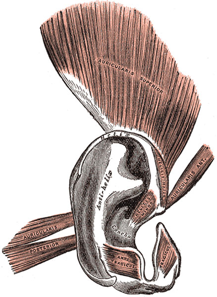
Auricula Muscles
Muscles
The muscles of the auricula (Fig. 906) consist of two sets: (1) the extrinsic, which connect it with the skull and scalp and move the auricula as a whole; and (2) the intrinsic, which extend from one part of the auricle to another.
The extrinsic muscles are the:
- Auricularis anterior - (Attrahens aurem), the smallest of the three, is thin, fan-shaped, and its fibers are pale and indistinct. It arises from the lateral edge of the galea aponeurotica, and its fibers converge to be inserted into a projection on the front of the helix.
- Auricularis superior - (Attolens aurem), the largest of the three, is thin and fan-shaped. Its fibers arise from the galea aponeurotica, and converge to be inserted by a thin, flattened tendon into the upper part of the cranial surface of the auricula.
- Auricularis posterior - (Retrahens aurem) consists of two or three fleshy fasciculi, which arise from the mastoid portion of the temporal bone by short aponeurotic fibers. They are inserted into the lower part of the cranial surface of the concha.
Actions.—In man, these muscles possess very little action: the Auricularis anterior draws the auricula forward and upward; the Auricularis superior slightly raises it; and the Auricularis posterior draws it backward.
The intrinsic muscles are the:
- Helicis major - is a narrow vertical band situated upon the anterior margin of the helix. It arises below, from the spina helicis, and is inserted into the anterior border of the helix, just where it is about to curve backward.
- Helicis minor - is an oblique fasciculus, covering the crus helicis.
- Tragicus - is a short, flattened vertical band on the lateral surface of the tragus.
- Antitragicus - arises from the outer part of the antitragus, and is inserted into the cauda helicis and antihelix.
- Transversus auriculæ - is placed on the cranial surface of the pinna. It consists of scattered fibers, partly tendinous and partly muscular, extending from the eminentia conchæ to the prominence corresponding with the scapha.
- Obliquus auriculæ - also on the cranial surface, consists of a few fibers extending from the upper and back part of the concha to the convexity immediately above it.
Nerves
- The Auriculares anterior and superior and the intrinsic muscles on the lateral surface are supplied by the temporal branch of the facial nerve.
- The Auricularis posterior and the intrinsic muscles on the cranial surface by the posterior auricular branch of the facial nerve.
Ligaments
The ligaments of the auricula (ligamenti auricularia [Valsalva]; ligaments of the pinna) consist of two sets:
- extrinsic, connecting it to the side of the head
- intrinsic, connecting various parts of its cartilage together.
The extrinsic ligaments are two in number, anterior and posterior. The anterior ligament extends from the tragus and spina helicis to the root of the zygomatic process of the temporal bone. The posterior ligament passes from the posterior surface of the concha to the outer surface of the mastoid process.
The chief intrinsic ligaments are: (a) a strong fibrous band, stretching from the tragus to the commencement of the helix, completing the meatus in front, and partly encircling the boundary of the concha; and (b) a band between the antihelix and the cauda helicis. Other less important bands are found on the cranial surface of the pinna.
Cartilage
The cartilage of the auricula (cartilago auriculæ; cartilage of the pinna) (Fig. 905, Fig. 906) consists of a single piece; it gives form to this part of the ear, and upon its surface are found the eminences and depressions above described.
It is absent from the lobule; it is deficient, also, between the tragus and beginning of the helix, the gap being filled up by dense fibrous tissue. At the front part of the auricula, where the helix bends upward, is a small projection of cartilage, called the spina helicis, while in the lower part of the helix the cartilage is prolonged downward as a tail-like process, the cauda helicis; this is separated from the antihelix by a fissure, the fissura antitragohelicina. The cranial aspect of the cartilage exhibits a transverse furrow, the sulcus antihelicis transversus, which corresponds with the inferior crus of the antihelix and separates the eminentia conchæ from the eminentia triangularis. The eminentia conchæ is crossed by a vertical ridge (ponticulus), which gives attachment to the Auricularis posterior muscle. In the cartilage of the auricula are two fissures, one behind the crus helicis and another in the tragus.
(Text modified from Gray's 1918 Anatomy)
- Gray's Images: Development | Lymphatic | Neural | Vision | Hearing | Somatosensory | Integumentary | Respiratory | Gastrointestinal | Urogenital | Endocrine | Surface Anatomy | iBook | Historic Disclaimer
| Historic Disclaimer - information about historic embryology pages |
|---|
| Pages where the terms "Historic" (textbooks, papers, people, recommendations) appear on this site, and sections within pages where this disclaimer appears, indicate that the content and scientific understanding are specific to the time of publication. This means that while some scientific descriptions are still accurate, the terminology and interpretation of the developmental mechanisms reflect the understanding at the time of original publication and those of the preceding periods, these terms, interpretations and recommendations may not reflect our current scientific understanding. (More? Embryology History | Historic Embryology Papers) |
| iBook - Gray's Embryology | |
|---|---|

|
|
Reference
Gray H. Anatomy of the human body. (1918) Philadelphia: Lea & Febiger.
Cite this page: Hill, M.A. (2024, April 27) Embryology Gray0906.jpg. Retrieved from https://embryology.med.unsw.edu.au/embryology/index.php/File:Gray0906.jpg
- © Dr Mark Hill 2024, UNSW Embryology ISBN: 978 0 7334 2609 4 - UNSW CRICOS Provider Code No. 00098G
File history
Click on a date/time to view the file as it appeared at that time.
| Date/Time | Thumbnail | Dimensions | User | Comment | |
|---|---|---|---|---|---|
| current | 06:13, 19 August 2012 |  | 438 × 600 (81 KB) | Z8600021 (talk | contribs) | ==Cartilage of Right Auricula== Cranial surface of cartilage of right auricula. The cartilage of the auricula (cartilago auriculæ; cartilage of the pinna) (Fig. 905, Fig. 906) consists of a single piece; it |
You cannot overwrite this file.
File usage
The following page uses this file:

