File:Gray0904.jpg
Gray0904.jpg (226 × 350 pixels, file size: 26 KB, MIME type: image/jpeg)
Auricula or Pinna
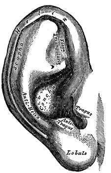
The auricula. Lateral surface
The external ear consists of the expanded portion named the auricula or pinna, and the external acoustic meatus. The former projects from the side of the head and serves to collect the vibrations of the air by which sound is produced; the latter leads inward from the bottom of the auricula and conducts the vibrations to the tympanic cavity.
The Auricula or Pinna (Fig. 904) is of an ovoid form, with its larger end directed upward. Its lateral surface is irregularly concave, directed slightly forward, and presents numerous eminences and depressions to which names have been assigned. The prominent rim of the auricula is called the helix; where the helix turns downward behind, a small tubercle, the auricular tubercle of Darwin, is frequently seen; this tubercle is very evident about the sixth month of fetal life when the whole auricula has a close resemblance to that of some of the adult monkeys. Another curved prominence, parallel with and in front of the helix, is called the antihelix; this divides above into two crura, between which is a triangular depression, the fossa triangularis. The narrow-curved depression between the helix and the antihelix is called the scapha; the antihelix describes a curve around a deep, capacious cavity, the concha, which is partially divided into two parts by the crus or commencement of the helix; the upper part is termed the cymba conchæ, the lower part the cavum conchæ. In front of the concha, and projecting backward over the meatus, is a small pointed eminence, the tragus, so called from its being generally covered on its under surface with a tuft of hair, resembling a goat’s beard. Opposite the tragus, and separated from it by the intertragic notch, is a small tubercle, the antitragus. Below this is the lobule, composed of tough areolar and adipose tissues, and wanting the firmness and elasticity of the rest of the auricula.
(Text modified from Gray's 1918 Anatomy)
- Gray's Images: Development | Lymphatic | Neural | Vision | Hearing | Somatosensory | Integumentary | Respiratory | Gastrointestinal | Urogenital | Endocrine | Surface Anatomy | iBook | Historic Disclaimer
| Historic Disclaimer - information about historic embryology pages |
|---|
| Pages where the terms "Historic" (textbooks, papers, people, recommendations) appear on this site, and sections within pages where this disclaimer appears, indicate that the content and scientific understanding are specific to the time of publication. This means that while some scientific descriptions are still accurate, the terminology and interpretation of the developmental mechanisms reflect the understanding at the time of original publication and those of the preceding periods, these terms, interpretations and recommendations may not reflect our current scientific understanding. (More? Embryology History | Historic Embryology Papers) |
| iBook - Gray's Embryology | |
|---|---|

|
|
Reference
Gray H. Anatomy of the human body. (1918) Philadelphia: Lea & Febiger.
Cite this page: Hill, M.A. (2024, April 27) Embryology Gray0904.jpg. Retrieved from https://embryology.med.unsw.edu.au/embryology/index.php/File:Gray0904.jpg
- © Dr Mark Hill 2024, UNSW Embryology ISBN: 978 0 7334 2609 4 - UNSW CRICOS Provider Code No. 00098G
File history
Click on a date/time to view the file as it appeared at that time.
| Date/Time | Thumbnail | Dimensions | User | Comment | |
|---|---|---|---|---|---|
| current | 14:48, 4 June 2010 |  | 226 × 350 (26 KB) | S8600021 (talk | contribs) | Category:Historic Embryology Category:Gray's 1918 Anatomy Category:Cartoon Category:Senses Category:Hearing |
You cannot overwrite this file.
File usage
The following 2 pages use this file:

