File:Gray0675.jpg
Gray0675.jpg (615 × 600 pixels, file size: 69 KB, MIME type: image/jpeg)
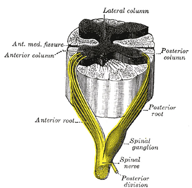
Fig. 675. A spinal nerve with its anterior and posterior roots
Roots of the Spinal Nerves
As already stated, each spinal nerve possesses two roots, an anterior and a posterior, which are attached to the surface of the medulla spinalis opposite the corresponding column of gray substance (Fig. 675); their fibers become medullated about the fifth month of fetal life.
Anterior Nerve Root
(radix anterior) Consists of efferent fibers, which are the axons of the nerve cells in the ventral part of the anterior and lateral columns. A short distance from their origins, these axons are invested by medullary sheaths and, passing forward, emerge in two or three irregular rows over an area which measures about 3 mm. in width.
Posterior Root
(radix posterior) Comprises some six or eight fasciculi, attached in linear series along the postero-lateral sulcus. It consists of afferent fibers which arise from the nerve cells in a spinal ganglion. Each ganglion cell gives off a single fiber which divides in a T-shaped manner into two processes, medial and lateral. The lateral processes extend to the sensory end-organs of the skin, muscles, tendons, joints, etc. (somatic receptors), and to the sensory end-organs of the viscera (visceral receptors). The medial processes of the ganglion cells grow into the medulla spinalis as the posterior roots of the spinal nerves.
- Gray's Images: Development | Lymphatic | Neural | Vision | Hearing | Somatosensory | Integumentary | Respiratory | Gastrointestinal | Urogenital | Endocrine | Surface Anatomy | iBook | Historic Disclaimer
| Historic Disclaimer - information about historic embryology pages |
|---|
 |
| iBook - Gray's Embryology | |
|---|---|

|
|
Reference
Gray H. Anatomy of the human body. (1918) Philadelphia: Lea & Febiger.
Cite this page: Hill, M.A. (2024, April 27) Embryology Gray0675.jpg. Retrieved from https://embryology.med.unsw.edu.au/embryology/index.php/File:Gray0675.jpg
- © Dr Mark Hill 2024, UNSW Embryology ISBN: 978 0 7334 2609 4 - UNSW CRICOS Provider Code No. 00098G
File history
Click on a date/time to view the file as it appeared at that time.
| Date/Time | Thumbnail | Dimensions | User | Comment | |
|---|---|---|---|---|---|
| current | 13:10, 17 October 2012 | | 615 × 600 (69 KB) | Z8600021 (talk | contribs) | ==Fig. 675. A spinal nerve with its anterior and posterior roots== Roots of the Spinal Nerves As already stated, each spinal nerve possesses two roots, an anterior and a posterior, which are attached to the surface of the medulla spinalis opposite the c |
You cannot overwrite this file.