File:Gray0658.jpg

Gray0658.jpg (361 × 450 pixels, file size: 29 KB, MIME type: image/jpeg)
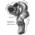
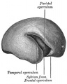
Human Fetal Brain (5 months)
Outer surface of cerebral hemisphere of human embryo of about five months.
- Brain Development Links: Week 4.5 exterior | Week 5 exterior | Week 5 interior | 3 month | 3 month hindbrain | 4 month | 5 month | Gray's Neural Images | Neural System Development
651 Human Embryo Brain (week 4.5 exterior view)
652 Human Embryo Brain (week 5 exterior view)
653 Human Embryo Brain (week 5 interior view)

654 Human Fetal Brain (3 months)
655 Human Fetal Brain (4 months)
658 Human Fetal Brain (5 months)


Fissures and Sulci
The outer surface of the cerebral hemisphere is at first smooth, but later it exhibits a number of elevations or convolutions, separated from each other by fissures and sulci, most of which make their appearance during the sixth or seventh months of fetal life.
The term fissure is applied to such grooves as involve the entire thickness of the cerebral wall, and thus produce corresponding eminences in the ventricular cavity, while the sulci affect only the superficial part of the wall, and therefore leave no impressions in the ventricle.
The fissures comprise the choroidal and hippocampal already described, and two others, viz., the calcarine and collateral, which produce the swellings known respectively as the calcar avis and the collateral eminence in the ventricular cavity. Of the sulci the following may be referred to, viz., the central sulcus (fissure of Rolando), which is developed in two parts; the intraparietal sulcus in four parts; and the cingulate sulcus in two or three parts.
The lateral cerebral or Sylvian fissure differs from all the other fissures in its mode of development. It appears about the third month as a depression, the Sylvian fossa, on the lateral surface of the hemisphere (Fig. 658); this fossa corresponds with the position of the corpus striatum, and its floor is moulded to form the insula. The intimate connection which exists between the cortex of the insula and the subjacent corpus striatum prevents this part of the hemisphere wall from expanding at the same rate as the portions which surround it. The neighboring parts of the hemisphere therefore gradually grow over and cover in the insula, and constitute the temporal, parietal, frontal, and orbital opercula of the adult brain. The frontal and orbital opercula are the last to form, but by the end of the first year after birth the insula is completely submerged by the approximation of the opercula. The fissures separating the opposed margins of the opercula constitute the composite lateral cerebral fissure.
- Gray's Images: Development | Lymphatic | Neural | Vision | Hearing | Somatosensory | Integumentary | Respiratory | Gastrointestinal | Urogenital | Endocrine | Surface Anatomy | iBook | Historic Disclaimer
| Historic Disclaimer - information about historic embryology pages |
|---|
 |
| iBook - Gray's Embryology | |
|---|---|

|
|
Reference
Gray H. Anatomy of the human body. (1918) Philadelphia: Lea & Febiger.
Cite this page: Hill, M.A. (2024, April 27) Embryology Gray0658.jpg. Retrieved from https://embryology.med.unsw.edu.au/embryology/index.php/File:Gray0658.jpg
- © Dr Mark Hill 2024, UNSW Embryology ISBN: 978 0 7334 2609 4 - UNSW CRICOS Provider Code No. 00098G
File history
Click on a date/time to view the file as it appeared at that time.
| Date/Time | Thumbnail | Dimensions | User | Comment | |
|---|---|---|---|---|---|
| current | 16:25, 11 October 2009 | | 361 × 450 (29 KB) | S8600021 (talk | contribs) | Outer surface of cerebral hemisphere of human embryo of about five months. Image Source: Anatomy of the Human Body (1918) by Henry Gray Category:Historic Embryology Category:Gray's 1918 Anatomy Category:Neural |
You cannot overwrite this file.
File usage
The following 18 pages use this file:
- 2009 Lecture 22
- 2010 Lab 12
- 2010 Lecture 23
- ANAT2341 Lab 10 - Fetal
- Anatomy of the Human Body by Henry Gray
- Lecture - Fetal Development
- Lecture - Neural Development
- Neural - Cerebrum Development
- Neural System - Fetal
- User:Z5019799
- File:Gray0649.jpg
- File:Gray0651.jpg
- File:Gray0652.jpg
- File:Gray0653.jpg
- File:Gray0654.jpg
- File:Gray0655.jpg
- File:Gray0658.jpg
- Template:Gray-Brain