File:Gray0654.jpg

Gray0654.jpg (402 × 500 pixels, file size: 39 KB, MIME type: image/jpeg)
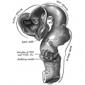
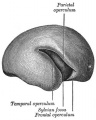
Human Fetal Brain (3 months)
Median sagittal section of brain of human embryo of three months. From model by Wilhelm His (1831-1904).
- Brain Development Links: Week 4.5 exterior | Week 5 exterior | Week 5 interior | 3 month | 3 month hindbrain | 4 month | 5 month | Gray's Neural Images | Neural System Development
651 Human Embryo Brain (week 4.5 exterior view)
652 Human Embryo Brain (week 5 exterior view)
653 Human Embryo Brain (week 5 interior view)

654 Human Fetal Brain (3 months)
655 Human Fetal Brain (4 months)
658 Human Fetal Brain (5 months)


The Diencephalon
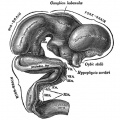
From the alar lamina of the diencephalon, the thalamus, metathalamus, and epithalamus are developed.
The thalamus (Figs. 650 to 654) arises as a thickening which involves the anterior two-thirds of the alar lamina. The two thalami are visible, for a time, on the surface of the brain, but are subsequently hidden by the cerebral hemispheres which grow backward over them. The thalami extend medialward and gradually narrow the cavity between them into a slit-like aperture which forms the greater part of the third ventricle; their medial surfaces ultimately adhere, in part, to each other, and the intermediate mass of the ventricle is developed across the area of contact.
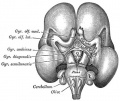
The metathalamus comprises the geniculate bodies which originate as slight outward bulgings of the alar lamina. In the adult the lateral geniculate body appears as an eminence on the lateral part of the posterior end of the thalamus, while the medial is situated on the lateral aspect of the mid-brain.
The epithalamus includes the pineal body, the posterior commissure, and the trigonum habenulæ.
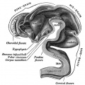
- The pineal body arises as an upward the evagination of roof-plate immediately in front of the midbrian; this evagination becomes solid with the exception of its proximal part, which persists as the recessus pinealis. In lizards the pineal evagination is elongated into a stalk, and its peripheral extremity is expanded into a vesicle, in which a rudimentary lens and retina are formed; the stalk becomes solid and nerve fibers make their appearance in it, so that in these animals the pineal body forms a rudimentary eye.
- The posterior commissure is formed by the ingrowth of fibers into the depression behind and below the pineal evagination, and the trigonum habenulæ is developed in front of the pineal recess.
- From the basal laminæ of the diencephalon the pars mamillaris hypothalami is developed; this comprises the corpora mamillaria and the posterior part of the tuber cinereum. The corpora mamillaria arise as a single thickening, which becomes divided into two by a median furrow during the third month. The roof-plate of the diencephalon, in front of the pineal body, remains thin and epithelial in character, and is subsequently invaginated by the choroid plexuses of the third ventricle.
- Gray's Images: Development | Lymphatic | Neural | Vision | Hearing | Somatosensory | Integumentary | Respiratory | Gastrointestinal | Urogenital | Endocrine | Surface Anatomy | iBook | Historic Disclaimer
| Historic Disclaimer - information about historic embryology pages |
|---|
 |
| iBook - Gray's Embryology | |
|---|---|

|
|
Reference
Gray H. Anatomy of the human body. (1918) Philadelphia: Lea & Febiger.
Cite this page: Hill, M.A. (2024, April 27) Embryology Gray0654.jpg. Retrieved from https://embryology.med.unsw.edu.au/embryology/index.php/File:Gray0654.jpg
- © Dr Mark Hill 2024, UNSW Embryology ISBN: 978 0 7334 2609 4 - UNSW CRICOS Provider Code No. 00098G
File history
Click on a date/time to view the file as it appeared at that time.
| Date/Time | Thumbnail | Dimensions | User | Comment | |
|---|---|---|---|---|---|
| current | 16:11, 11 October 2009 | | 402 × 500 (39 KB) | S8600021 (talk | contribs) | Median sagittal section of brain of human embryo of three months. (From model by His.) Image Source: Anatomy of the Human Body (1918) by Henry Gray Category:Historic Embryology Category:Gray's 1918 Anatomy Category:Neural |
You cannot overwrite this file.
File usage
The following 24 pages use this file:
- 2009 Lecture 22
- 2010 Lab 12
- 2010 Lecture 23
- ANAT2341 Lab 10 - Fetal
- Anatomy of the Human Body by Henry Gray
- BGD Lecture - Endocrine Development
- Endocrine - Hypothalamus Development
- Lecture - Endocrine Development
- Lecture - Fetal Development
- Lecture - Neural Development
- Neural - Cerebrum Development
- Neural - Thalamus Development
- Neural System - Fetal
- REI - Reproductive Medicine Seminar 2018
- Royal Hospital for Women - Reproductive Medicine Seminar 2018
- User:Z5019799
- File:Gray0649.jpg
- File:Gray0651.jpg
- File:Gray0652.jpg
- File:Gray0653.jpg
- File:Gray0654.jpg
- File:Gray0655.jpg
- File:Gray0658.jpg
- Template:Gray-Brain