File:Gray0649.jpg

Original file (698 × 700 pixels, file size: 72 KB, MIME type: image/jpeg)
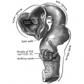
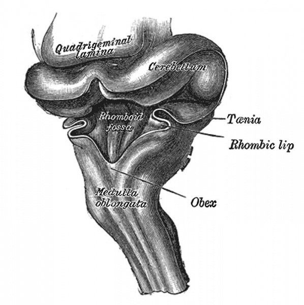
Hind-brain of a human embryo of three months
Viewed from behind and partly from left side. (From model by His.)
The Hind-brain or Rhombencephalon
The cavity of the hind-brain becomes the fourth ventricle. At the time when the ventral cephalic flexure makes its appearance, the length of the hind-brain exceeds the combined lengths of the other two vesicles. Immediately behind the mid-brain it exhibits a marked constriction, the isthmus rhombencephali (Fig. 650, Isthmus), which is best seen when the brain is viewed from the dorsal aspect. From the isthmus the anterior medullary velum and the superior peduncle of the cerebellum are formed. It is customary to divide the rest of the hind-brain into two parts, viz., an upper, called the metencephalon, and a lower, the myelencephalon. The cerebellum is developed by a thickening of the roof, and the pons by a thickening in the floor and lateral walls of the metencephalon. The floor and lateral walls of the myelencephalon are thickened to form the medulla oblongata; its roof remains thin, and, retaining to a great extent its epithelial nature, is expanded in a lateral direction. Later, by the growth and backward extension of the cerebellum, the roof is folded inward toward the cavity of the fourth ventricle; it assists in completing the dorsal wall of this cavity, and is also invaginated to form the ependymal covering of its choroid plexuses. Above it is continuous with the posterior medullary velum; below, with the obex and ligulæ.
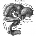
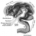
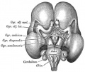
- Brain Development Links: Week 4.5 exterior | Week 5 exterior | Week 5 interior | 3 month | 3 month hindbrain | 4 month | 5 month | Gray's Neural Images | Neural System Development
651 Human Embryo Brain (week 4.5 exterior view)
652 Human Embryo Brain (week 5 exterior view)
653 Human Embryo Brain (week 5 interior view)

654 Human Fetal Brain (3 months)
655 Human Fetal Brain (4 months)
658 Human Fetal Brain (5 months)


- Gray's Images: Development | Lymphatic | Neural | Vision | Hearing | Somatosensory | Integumentary | Respiratory | Gastrointestinal | Urogenital | Endocrine | Surface Anatomy | iBook | Historic Disclaimer
| Historic Disclaimer - information about historic embryology pages |
|---|
 |
| iBook - Gray's Embryology | |
|---|---|

|
|
Reference
Gray H. Anatomy of the human body. (1918) Philadelphia: Lea & Febiger.
Cite this page: Hill, M.A. (2024, April 27) Embryology Gray0649.jpg. Retrieved from https://embryology.med.unsw.edu.au/embryology/index.php/File:Gray0649.jpg
- © Dr Mark Hill 2024, UNSW Embryology ISBN: 978 0 7334 2609 4 - UNSW CRICOS Provider Code No. 00098G
File history
Click on a date/time to view the file as it appeared at that time.
| Date/Time | Thumbnail | Dimensions | User | Comment | |
|---|---|---|---|---|---|
| current | 09:04, 20 May 2012 | | 698 × 700 (72 KB) | Z8600021 (talk | contribs) |
You cannot overwrite this file.
File usage
The following page uses this file: