File:Gillilan1959-fig03.jpg
Original file (1,042 × 1,366 pixels, file size: 317 KB, MIME type: image/jpeg)
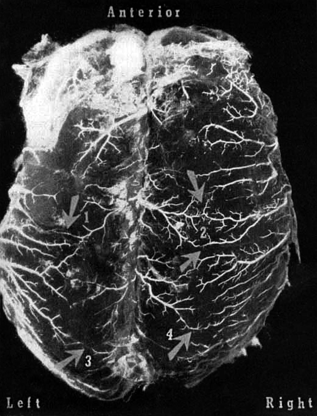
Fig. 3. Fetal Brain (6 month) Large Superficial Arteries
A photograph of a 6-months old fetal brain, with the large superficial arteries injected, and the brain cleared by the Spalteholz technique.
Arrows 1 and 2 indicate anastomoses between large branches of the anterior cerebral arteries and the middle cerebral arteries.
Arrows 3 and 4 point out anastomoses between the posterior cerebral and the middle cerebral arteries. These large superficial anastomoses can be seen clearly only before the development of the gyri and sulci.
| Historic Disclaimer - information about historic embryology pages |
|---|
 |
Reference
Gillilan, LA. Significant superficial anastomoses in the arterial blood supply to the human brain. J Comp Neurol. 1959 Jun;112:55-74. PMID 13850118
File history
Click on a date/time to view the file as it appeared at that time.
| Date/Time | Thumbnail | Dimensions | User | Comment | |
|---|---|---|---|---|---|
| current | 10:01, 16 November 2015 | | 1,042 × 1,366 (317 KB) | Z8600021 (talk | contribs) | |
| 10:01, 16 November 2015 | 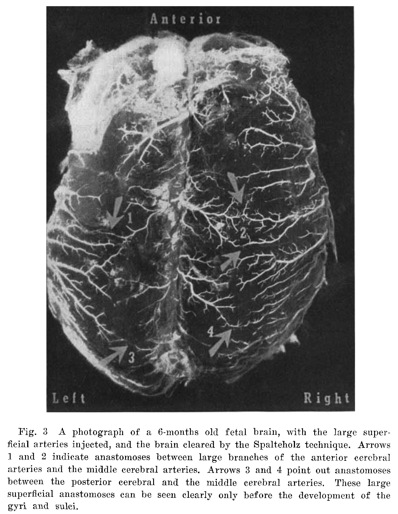 | 1,349 × 1,739 (367 KB) | Z8600021 (talk | contribs) |
You cannot overwrite this file.
File usage
The following 2 pages use this file: