File:Gillilan1959-fig02.jpg
Original file (905 × 1,000 pixels, file size: 159 KB, MIME type: image/jpeg)
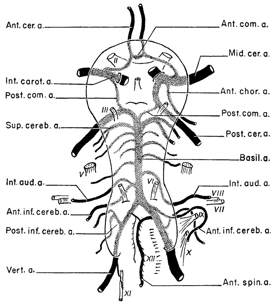
Fig. 2. Complex of arteries on the Ventral Surface of the Brain
| A diagram illustrating the complex of arteries seen on the ventral surface of the brain.
The arteries drawn in solid black represent the 4 large arteries supplying the brain and the constant group of small arteries supplying specific areas of the brain. The arteries lying inside the elliptoid line and which are stippled represent the highly variable, medium-sized group. Eighty-five % of cerebral aneurysms are located inside the elliptoid line, occurring on the variable group of arteries.
|
ReferenceGillilan, LA. Significant superficial anastomoses in the arterial blood supply to the human brain. J Comp Neurol. 1959 Jun;112:55-74. PMID 13850118[ |

File history
Click on a date/time to view the file as it appeared at that time.
| Date/Time | Thumbnail | Dimensions | User | Comment | |
|---|---|---|---|---|---|
| current | 10:17, 16 November 2015 | | 905 × 1,000 (159 KB) | Z8600021 (talk | contribs) | |
| 10:16, 16 November 2015 | 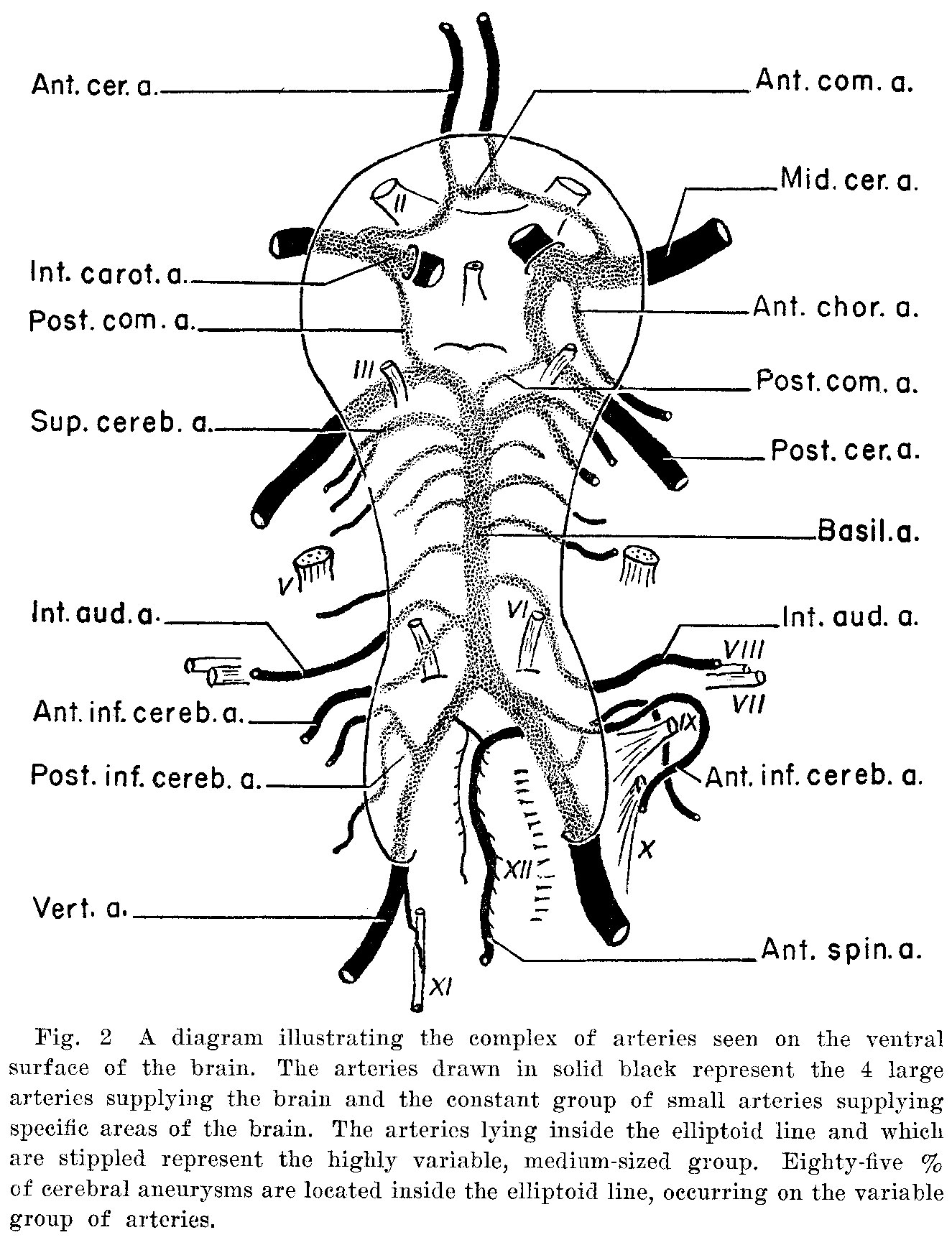 | 1,325 × 1,726 (408 KB) | Z8600021 (talk | contribs) | {{Historic Disclaimer}} Historic Embryology Papers ===Reference=== Gillilan, LA. [[Paper - Significant superficial anastomoses in the arterial blood supply to the human brain|Significant superficial anastomoses in the arterial blood supply to the... |
You cannot overwrite this file.
File usage
The following 2 pages use this file: