File:Gap junction 01.jpg
From Embryology
No higher resolution available.
Gap_junction_01.jpg (800 × 562 pixels, file size: 69 KB, MIME type: image/jpeg)
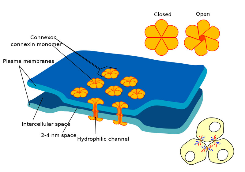
Gap Junctions
Discovered by J.P. Revel & M.J. Karnovsky in 1967
- allowing direct communication between cells (open & close)
- close membranes 2 - 3 nm apart
- connexins form hollow 1.5 nm diameter cylinders
- heart muscle, smooth muscle electrical and chemical integration as a single functional unit
- Also in embryonic development (see Blastocyst Development)
Search PubMed: gap junction
- Adhesion EM Images: GIT epithelia EM1 | GIT epithelia EM2 | GIT epithelia EM3 | Desmosome EM
- Adhesion Cartoons: Tight junction | Adherens Junction | Desmosome | Gap Junction
Cite this page: Hill, M.A. (2024, April 27) Embryology Gap junction 01.jpg. Retrieved from https://embryology.med.unsw.edu.au/embryology/index.php/File:Gap_junction_01.jpg
- © Dr Mark Hill 2024, UNSW Embryology ISBN: 978 0 7334 2609 4 - UNSW CRICOS Provider Code No. 00098G
File history
Click on a date/time to view the file as it appeared at that time.
| Date/Time | Thumbnail | Dimensions | User | Comment | |
|---|---|---|---|---|---|
| current | 12:15, 3 March 2012 | | 800 × 562 (69 KB) | Z8600021 (talk | contribs) | ==Gap Junctions== * Discovered by J.P. Revel & M.J. Karnovsky in 1967 * allowing direct communication between cells (open & close) * close membranes 2 - 3 nm apart * connexins form hollow 1.5 nm diameter cylinders * heart muscle, smooth muscle electrical |
You cannot overwrite this file.