File:Frazer1928 fig02.jpg: Difference between revisions
mNo edit summary |
mNo edit summary |
||
| (One intermediate revision by the same user not shown) | |||
| Line 1: | Line 1: | ||
==Text-fig. 2. On the left, a scheme showing the component parts in the regions of hind-brain== | ==Text-fig. 2. On the left, a scheme showing the component parts in the regions of hind-brain 12 mm Embryo== | ||
H ., and mid-brain, M. Further explanation in text. The arrow gives direction of section (on right). BL. basal lamina, FL. floor lamina, 221.3. intralaminar sulcus, a. f. area. of short association fibres. | H ., and mid-brain, M. Further explanation in text. The arrow gives direction of section (on right). BL. basal lamina, FL. floor lamina, 221.3. intralaminar sulcus, a. f. area. of short association fibres. | ||
The first figure in text-fig. 2 is a diagram intended to show the disposition of the several parts entering into the formation of the wall of the cavity in the 12 mm. embryo. In the hind-brain (H) the basal and alar areas are represented respectively by cross-hatching and small circles, while the tectal region of the mid-brain (M) is shown by dots. The floor lamina of the mid-brain is marked like the basal lamina, with which it is continuous. It must be noted here that this floor lamina contains a Well-developed zone of short association fibres lying against its thick ependymal layer, extending back to the basal lamina and forward to the region of the third nerve nucleus. | |||
The transverse section (text-fig. 2) thus shows a lower part, the floor lamina, marked by the association fibres and separated above from the upper part by a deep cleft, which is the intralaminar sulcus. The part above this sulcus is the basal lamina, containing no association fibres. The trochlear nerve, issuing from the hind-brain at this stage, is not cut at the top of the section, but is divided lower down near its nucleus, which is just in front of the line of section. Thus there are nothing but basal and floor structures in the section of the isthmus region, and the thickness of the floor lamina seems to be due almost entirely to the large formation of association fibres. | |||
Latest revision as of 13:19, 9 January 2017
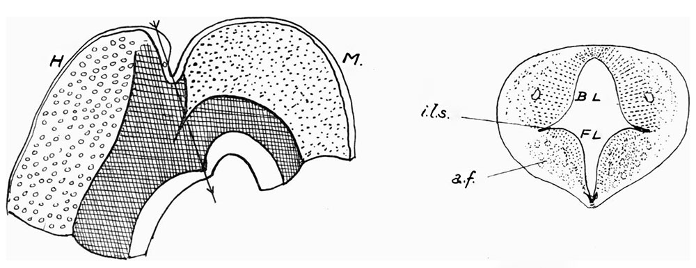
Text-fig. 2. On the left, a scheme showing the component parts in the regions of hind-brain 12 mm Embryo
H ., and mid-brain, M. Further explanation in text. The arrow gives direction of section (on right). BL. basal lamina, FL. floor lamina, 221.3. intralaminar sulcus, a. f. area. of short association fibres.
The first figure in text-fig. 2 is a diagram intended to show the disposition of the several parts entering into the formation of the wall of the cavity in the 12 mm. embryo. In the hind-brain (H) the basal and alar areas are represented respectively by cross-hatching and small circles, while the tectal region of the mid-brain (M) is shown by dots. The floor lamina of the mid-brain is marked like the basal lamina, with which it is continuous. It must be noted here that this floor lamina contains a Well-developed zone of short association fibres lying against its thick ependymal layer, extending back to the basal lamina and forward to the region of the third nerve nucleus.
The transverse section (text-fig. 2) thus shows a lower part, the floor lamina, marked by the association fibres and separated above from the upper part by a deep cleft, which is the intralaminar sulcus. The part above this sulcus is the basal lamina, containing no association fibres. The trochlear nerve, issuing from the hind-brain at this stage, is not cut at the top of the section, but is divided lower down near its nucleus, which is just in front of the line of section. Thus there are nothing but basal and floor structures in the section of the isthmus region, and the thickness of the floor lamina seems to be due almost entirely to the large formation of association fibres.
| Historic Disclaimer - information about historic embryology pages |
|---|
 |
Reference
Frazer JE. Development of the region of the isthmus rhombencephali. (1928) J Anat. 63: 7-18. PMID 17104212
Cite this page: Hill, M.A. (2024, April 27) Embryology Frazer1928 fig02.jpg. Retrieved from https://embryology.med.unsw.edu.au/embryology/index.php/File:Frazer1928_fig02.jpg
- © Dr Mark Hill 2024, UNSW Embryology ISBN: 978 0 7334 2609 4 - UNSW CRICOS Provider Code No. 00098G
File history
Click on a date/time to view the file as it appeared at that time.
| Date/Time | Thumbnail | Dimensions | User | Comment | |
|---|---|---|---|---|---|
| current | 13:12, 9 January 2017 | 1,000 × 386 (84 KB) | Z8600021 (talk | contribs) | ||
| 13:10, 9 January 2017 |  | 1,451 × 711 (174 KB) | Z8600021 (talk | contribs) | {{Historic Disclaimer}} ===Reference=== {{Ref-Frazer1928}} {{Footer}} |
You cannot overwrite this file.
File usage
The following page uses this file: