File:Frazer1928 fig01.jpg
Original file (900 × 455 pixels, file size: 44 KB, MIME type: image/jpeg)
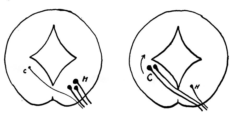
Text-fig. 1. Schemes to show the essentially similar nature of a dorsal efferent root and of the fourth nerve (right)
C., H. contralateral and homolateral origins. The arrow indicates that the contralateral origin of the fourth nerve is secondarily displaced further away, as is mentioned in the text.
The question of the meaning of the decussation after origin does not admit of solution on mechanical lines similar to the previous one, but there is, I think, good reason for saying that it may be explained on general grounds connected with the origin of many, if not all, efferent roots. It is known that the anterior roots of the spinal nerves, in addition to the fibres derived from ventral horn cells of their own side, possess also fibres which arise from cells situated on the other side of the cord. These contralateral fibres cross in the White ventral commissure and pass from there directly into the issuing root. The cells giving origin to them are described by Winkler as being in what he terms the “pars intermedia” of the cord, and the fibres are destined for the “sympathetic” system. Presumably, also, there is some cerebellar influence exercised on these cells, for Winkler describes cerebello-fugal fibres as reaching this same zone. We may say, then, that (in the dorsal, dorso-lumbar, and probably sacral regions at any rate) the spinal efferent roots have homolateral and contralateral origins: this is shown in the left-hand diagram in text-fig. 1, where C is the relatively small contralateral element in the root, and H the large and overshadowing homolateral portion. The condition which, it seemed to me, might be represented by the fourth nerve is shown on the right side, where the homolateral element is reduced to a minimum, while the contralateral portion is forming practically the whole of the nerve. This puts this nerve quite in line with the other, only differing in the proportions existing between its constituent parts, while its contralateral fibres cross in the ventral commissure, which is of course superficial here owing to the absence of the ventral projections which are so marked in the spinal cord. Taking this idea as a working hypothesis, a search was made for some representative of the homolateral origin, if perchance it might still be in evidence, and I think I have been able to find such a remnant: this has so far only been found in the older specimens, the younger ones being indeterminate in all their nuclear structures, as already pointed out.
| Historic Disclaimer - information about historic embryology pages |
|---|
 |
Reference
Frazer JE. Development of the region of the isthmus rhombencephali. (1928) J Anat. 63: 7-18. PMID 17104212
Cite this page: Hill, M.A. (2024, April 28) Embryology Frazer1928 fig01.jpg. Retrieved from https://embryology.med.unsw.edu.au/embryology/index.php/File:Frazer1928_fig01.jpg
- © Dr Mark Hill 2024, UNSW Embryology ISBN: 978 0 7334 2609 4 - UNSW CRICOS Provider Code No. 00098G
File history
Click on a date/time to view the file as it appeared at that time.
| Date/Time | Thumbnail | Dimensions | User | Comment | |
|---|---|---|---|---|---|
| current | 13:12, 9 January 2017 | | 900 × 455 (44 KB) | Z8600021 (talk | contribs) | |
| 13:10, 9 January 2017 | 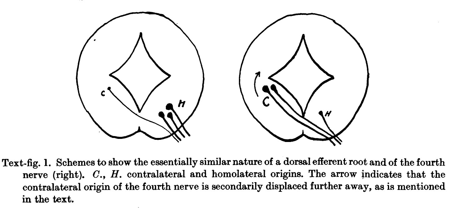 | 1,441 × 668 (120 KB) | Z8600021 (talk | contribs) | Text-fig. 1. Schemes to show the essentially similar nature of a dorsal efferent root and of the fourth nerve (right). 0., H. contralateral and homolateral origins. The arrow indicates that the contralateral origin of the fourth nerve is secondarily d... |
You cannot overwrite this file.
File usage
The following page uses this file: