File:Frazer1914 fig03.jpg

Original file (1,000 × 1,016 pixels, file size: 217 KB, MIME type: image/jpeg)
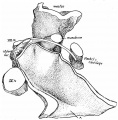
Fig. 3. Left recess from a model 27 mm Embryo
Observe that the forward extension, A, from third arch along the floor is much more marked and prominent than in the last figure, and has reached the first arch.
The further development of the region up to the third month is a simple progression of the changes seen in their early stages in fig. 2. The first arch area stands up more, the manubrial district of the second arch is more prominent and defined, and the second pouch is recognisable between the styloid bar and the glosso-pharyngeal nerve. But a distinct change is visible when attention is directed to the third arch region; the arch, presumably as" a result of its growth, is encroaching on the lumen of the inner portion of the cavity from behind, so that already some indication of a division into tubal and tympanic parts is foreshadowed. A drawing of one of the models showing this stage is seen in fig. 3, and it is interesting to compare it with the earlier condition: recognition of the first and second arch region is easy, and the districts into which the latter is subdivided are evident and unmistakable.
| Historic Disclaimer - information about historic embryology pages |
|---|
| Pages where the terms "Historic" (textbooks, papers, people, recommendations) appear on this site, and sections within pages where this disclaimer appears, indicate that the content and scientific understanding are specific to the time of publication. This means that while some scientific descriptions are still accurate, the terminology and interpretation of the developmental mechanisms reflect the understanding at the time of original publication and those of the preceding periods, these terms, interpretations and recommendations may not reflect our current scientific understanding. (More? Embryology History | Historic Embryology Papers) |
- Links: Fig 1 | Fig 2 | Fig 3 | Fig 4 | Fig 5 | Fig 6 | 1914 Frazer | Pharyngeal arches | Historic Embryology Papers
Reference
Frazer JE. The second visceral arch and groove in the tubo-tympanic region. (1914) J Anat Physiol. 48(4): 391-408. PMID 17233005
Cite this page: Hill, M.A. (2024, April 27) Embryology Frazer1914 fig03.jpg. Retrieved from https://embryology.med.unsw.edu.au/embryology/index.php/File:Frazer1914_fig03.jpg
- © Dr Mark Hill 2024, UNSW Embryology ISBN: 978 0 7334 2609 4 - UNSW CRICOS Provider Code No. 00098G
File history
Click on a date/time to view the file as it appeared at that time.
| Date/Time | Thumbnail | Dimensions | User | Comment | |
|---|---|---|---|---|---|
| current | 06:09, 9 January 2017 |  | 1,000 × 1,016 (217 KB) | Z8600021 (talk | contribs) | |
| 06:08, 9 January 2017 |  | 1,254 × 1,320 (277 KB) | Z8600021 (talk | contribs) |
You cannot overwrite this file.
File usage
The following 3 pages use this file:
